A new CT technique can measure blood vessel inflammation, allowing doctors to identify and treat patients at risk for cardiovascular disease, according to a U.K. study published online 12 July in Science Translational Medicine.
Cardiovascular disease accounts for one in every four deaths in the U.S., but the standard of care for assessing it -- coronary calcium scoring -- identifies hardened arteries when the damage has already become irreversible, and it can't distinguish which blood vessel plaques are prone to rupture.
A team led by Dr. Alexios Antonopoulos, PhD, of the University of Oxford created a method to identify blood vessel inflammation from measurements of fat tissue around arteries using CT. The technique uses a "fat attenuation index" (FAI), which is based on changes in fat cell size that correlated with markers of blood vessel inflammation in samples from 453 patients undergoing cardiac surgery. Among these patients, increased FAI correlated with blood vessel inflammation in a subset of 40 patients who were also evaluated with PET imaging.
Antonopoulos' group went on to quantify FAI in 273 additional subjects. They found that the marker changed around suspicious lesions in five patients who experienced heart attacks.
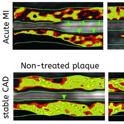
 A new imaging methodology called FAI identifies inflamed artery lesions that pose the most risk to patients. Image courtesy of Antonopoulos et al and Science Translational Medicine.
If ongoing follow-up studies confirm the prognostic value of FAI, the metric could transform risk stratification and clinical management for heart disease, according to the researchers.