Dear AuntMinnie Member,
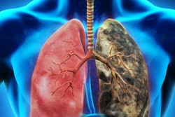
A new study published last week in BMJ found that the benefits of CT lung cancer screening in terms of reducing deaths from lung cancer far outweigh any iatrogenic cancers that might be caused by radiation from the scans.
The findings might not surprise anyone in radiology, but they are worth noting, in particular because they are published in a journal -- BMJ -- that historically has often taken a pretty skeptical view of medical imaging, especially when it's used for screening.
In the new study, researchers from Italy examined data from a 10-year CT lung screening study of more than 5,000 high-risk individuals. They compared the number of cancers found with how many cancers might be induced from 10 years of radiation delivered by the scans, based on statistical models.
Their findings? One radiation-induced cancer could be expected for every 108 lung cancers found, a trade-off that the group deemed acceptable. Learn more about the study by clicking here, or visit our CT Community at ct.auntminnie.com.
MRI of autism
In other news, visit our MRI Community for a new study in which researchers from North Carolina used MRI to detect what could be biomarkers indicating that infants might be at risk of developing autism in the future. The scans found 80% of the babies who would develop autism by age 2. Find out more by clicking here.
Also, learn about a new start-up company developing an MRI contrast technology that it hopes will work better than PET in detecting early signs of Alzheimer's disease. That story is available by clicking here, or visit our MRI Community at mri.auntminnie.com.
Breast density measurements
Finally, be sure to visit our Women's Imaging Community for two important new stories. First, researchers from California compared breast density measurements made by radiologists using the BI-RADS scale with those computed using automated software. Second, New York researchers found wide variations in how digital breast tomosynthesis has been implemented across the U.S.