Thursday, December 1 | All day | PH107-ED-X | Lakeside, PH Community
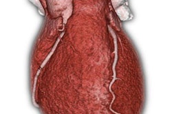
Can size-specific dose estimates (SSDEs) be considered the preferred dose index for detectability in lung cancer screening? This collaborative exhibit between Japanese and U.S. researchers explores the suitability of SSDE for dose assessment in lung cancer screening.As Japan deploys lung cancer screening with low-dose CT nationwide, its investigators are building a national registry to record both image quality and dose index values, explained Michael McNitt-Gray, PhD, from the University of California, Los Angeles.
The exhibit aims to report on image quality and radiation dose index values being collected in the registry, as well as relationships between the various index values and between dose index values and image quality, he stated. Specifically, the investigators examined CT dose index volume (CTDIvol), SSDE, and organ doses using Monte Carlo simulations, McNitt-Gray said.
"They also investigated the image noise and contrast-to-noise ratio using phantom scans," he said. "The data for each value and the trade-offs between these values will be presented. The clinical implications will be to first collect data and then advise sites carrying out low-dose lung cancer screening in Japan about the range of technical factors that provide adequate image quality for screening evaluations."