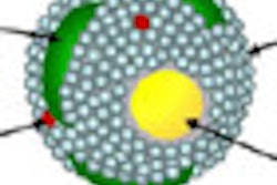
Texas researchers have succeeded in trapping bismuth in a nanotube cage, and the resultant structure could be used as a CT contrast agent to track stem cells, according to a new study in the Journal of Materials Chemistry B.
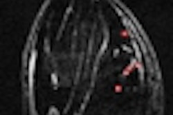
Specifically, the investigators from Rice University, in cooperation with the University of Houston and St. Luke's Episcopal Hospital, are inserting bismuth compounds into single-walled carbon nanotubes to make a more effective CT contrast agent. In tests using pig bone marrow-derived mesenchymal stem cells, the researchers found that the bismuth-filled nanotubes, which they have dubbed Bi@US-tubes, produce CT images of higher attenuation than those with iodine-based contrast agents (J Mater Chem B, October 7, 2013, Vol. 1:37, pp. 4792-4800).
Bismuth has been used before as a contrast agent, but putting it in nanotube capsules allowed the researchers to get the substance inside cells in high concentrations, permitting the acquisition of CT images of the cell. The relatively high contrast is achieved with low bismuth loading (2.66% by weight) within the tubes, without compromising cell viability.
Bismuth is a heavy element and therefore is more effective at diffracting x-rays than almost any substance, according to study co-author Lon Wilson, PhD. Going forward, the nanotube surfaces can be modified to improve biocompatibility and their ability to target certain types of cells. They can also be modified for use with MRI, PET, and electron paramagnetic resonance imaging systems, he said.
The researchers are now working to double the amount of bismuth in each nanotube. They would also like to combine bismuth and gadolinium into a single nanotube to produce a bimodal contrast agent suitable for tracking in both CT and MRI, Wilson said.
The research was supported by the Robert A. Welch Foundation, the U.S. National Institutes of Health, and the U.S. National Science Foundation.