Dual-energy CT accurately identifies anterior cruciate ligament (ACL) tears in the emergency setting, clearing a path to faster diagnosis of this common knee injury, investigators reported at this week's American Roentgen Ray Society (ARRS) meeting in Washington, DC.
In more than 50 patients, dual-energy CT showed greater than 90% sensitivity and similar specificity for detecting ACL tears in the emergency room (ER), said lead author Dr. Katrina Glazebrook from the Mayo Clinic in Rochester, MN.
Using dual-energy CT to identify significant internal derangement of the knee early can facilitate treatment planning for patients with knee trauma, she said in a statement.
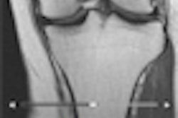
ACL tears are a common ligamentous injury of the knee, but they are rarely diagnosed in the emergency department because they are not seen on plain x-rays. MRI is the gold standard, but the exams can take upward of 40 minutes, and scanners are rarely sited in the emergency department; at the same time, CT scanners are more common in the ER setting.
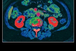
The team examined the knees of 27 patients using dual-energy CT in three planes: axial, sagittal, and oblique sagittal images to which a bone-removal algorithm and tendon-specific color mapping had been applied. The results showed that 16 patients had confirmed ACL tears, while 11 others had no history of trauma.
Two experienced readers, a musculoskeletal radiologist and a senior radiology resident, reviewed all the images. The radiologist achieved a 94% accuracy rate in identifying tears versus 87% for the resident.
This is a new use for dual-energy CT, but the images had sufficient spatial resolution and diagnostic quality to be interpreted by a physician not specializing in musculoskeletal trauma, the researchers wrote.