Today's cardiac CT is more robust than ever, broadening the range of patients who can be imaged. One new tool improves all-important temporal resolution even in midrange scanners. The software creates a hybrid sample of image data to improve temporal resolution in cardiac CT angiography (CCTA) images of obese patients.
"The main performance index of a cardiac CT study remains the temporal resolution of a fast acquisition," said Dr. Joseph Schoepf, a professor of cardiology and radiology at the Medical University of South Carolina. "If you have fast temporal resolution, your chances of catching a coronary artery within a state of quiescence are much better. Temporal resolution is the way you make sure you are ending up with high-quality images."
Schoepf spoke on the topic in June at the International Society for Computed Tomography (ISCT) meeting in San Francisco.
One way to achieve high temporal resolution is simply to buy an expensive scanner, he said. With the latest second-generation dual-source CT machines, for example, "you can zoom through the entire coronary artery tree" with an ultrafast scan of 270 msec acquired in less than a heartbeat. It's enough to cover a single diastole in a young patient, for example, at 75-msec temporal resolution, ruling out coronary artery disease with a radiation exposure of less than 1 mSv.
 |
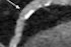
| Dual-source CT was able to rule out coronary artery disease in a 31-year-old man with chest pain and an abnormal SPECT exam. A 270-msec scan covered a single diastole at 75-msec temporal resolution with a radiation exposure of less than 1 mSv. All images courtesy of Dr. Joseph Schoepf. |
But not everyone can or will buy a high-end scanner, Schoepf noted. "Maybe your institution does hearts occasionally, but it doesn't constitute the brunt of your business, so a midrange scanner with limited gantry rotation speed is one of those scenarios," he said.
For these situations, and even for institutions with high-end scanners, a software application known as iTRIM (iterative temporal resolution improvement method) from Siemens Healthcare may be a good solution, he said.
Other techniques can also be used but don't measure up, according to Schoepf. For example, facilities with a midrange machine can maximize tube current and minimize scan range to defeat noise, but the lower noise comes at a price. "You obviously improve the photon flux through your obese patients, but the penalty is decreased temporal resolution if you artificially curb the gantry rotation speed of your scanner," he said.
Another potential solution is creating old-fashioned multisegment reconstructions, but this technique is also suboptimal because coronary arteries don't return to precisely the same location after successive heartbeats. The result is better temporal resolution, but also some inherent "inaccuracy and fuzziness" when reconstructing from such data, Schoepf said.
Recently, Schoepf's facility looked into a solution that "plays with some of the different compartments that you have in your CT data," he said. "The iTRIM algorithm is mainly aimed at enabling high-temporal-resolution coronary CT angiography on entry-level and midrange CT systems that have slower gantry rotation speeds. However, on all systems it can be used to mitigate the traditional trade-off between temporal resolution and image noise when imaging obese patients."
With iTRIM, the usual half-gantry rotation projection data are used to form a low-temporal-resolution but high-quality image that depicts structures outside of the heart, and also provides excellent contrast-to-noise ratio, Schoepf said. The algorithm is based on the finding that the presence of motion artifacts does not significantly change the histogram of a CT image.
iTRIM begins the process by creating a cardiac short-scan weighted filtered back projection (WFBP) reconstruction, resulting in a temporal resolution equivalent to 180º of rebinned CT data, according to a white paper from Siemens. For each pixel, the system computes a histogram within a quadratic region centered on the pixel. Next, an iterative reconstruction algorithm using a 140° subset of the full 180° set of cardiac data is adapted to the target temporal resolution. The low-temporal-resolution filtered back projection image is used as a start image for this iterative algorithm.
"You grab a subset of data -- not a 180º rotation but a 140º rotation -- and you send that through some iterative loops with that partial set of data that is kept in shape by the constraint image," Schoepf explained.
The two-step iterative loop first updates the projection data subset using the SART (simultaneous algebraic reconstruction technique) iterative reconstruction framework, according to the Siemens document. Then, after each SART iteration, the Hounsfield unit values of each pixel are adjusted according to the respective histogram of the low-temporal-resolution image. Pixels with HU values close to a maximum of the histogram are left unchanged, while pixels with values far less than the maximum are adjusted slightly toward the closest maximum.
"At the end of the process, you construct a hybrid image that consists of portions of the high-quality, low-temporal-resolution image and the high-temporal-resolution image," Schoepf explained. "You get the best of both worlds -- a high-resolution 120º image despite the use of rather slow gantry rotation speeds."
 |
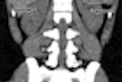
| Compared to filtered back projection reconstruction (above), use of the iTRIM algorithm improves temporal resolution and renders a more diagnostic image. |
 |
Testing iTRIM image quality
Schoepf and colleagues applied the iTRIM algorithm to a group of obese patients -- all had a body mass index (BMI) greater than 30 -- and compared the image quality of traditional FBP reconstruction at 250 msec temporal resolution with the iTRIM technique "where precious seconds were shaved off the temporal resolution to 200 msec," he said.
Results were as follows:
- There were no significant differences in attenuation between FBP- and TRIM-reconstructed series.
- There were no differences in contrast-to-noise ratio between FBP- and TRIM-reconstructed series.
- TRIM-reconstructed data showed significantly lower motion artifact severity scores than FBP (2.0 versus 2.5; p < 0.05).
 |
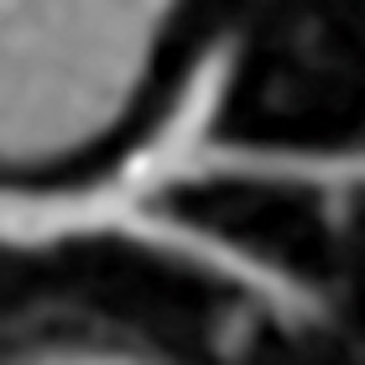
| Compared to filtered back projection reconstructions in CCTA of obese patients, subjective image quality was improved with use of the iTRIM algorithm. |
"This algorithm for cardiac CT allows you to obtain high-temporal-resolution cardiac CT images even if you're restricted by a midrange scan system, or if you want to artificially curb the gantry rotation speed because you're looking at obese individuals," Schoepf said.
Use of the algorithm mitigates the trade-off between temporal resolution and image noise when imaging obese patients, and it might improve the negative predictive value of slower gantry rotation systems to rule out significant coronary artery stenosis with coronary CTA, Schoepf said.
iTRIM has been cleared by the U.S. Food and Drug Administration (FDA) and is included in the toolkit of Siemens' new Somatom Perspective 128-detector-row CT scanner.