Dear CT Insider,
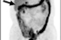
Among their many uses, iodine maps in dual-energy CT are a helpful way to gauge the size of lung perfusion defects in pulmonary embolism. But what's the information good for? No less than predicting patient outcomes, according to a research team from Germany and South Carolina.
The group compared perfusion defect size to several surrogate markers of pulmonary embolism severity including D-dimer levels, blood gas markers, and right heart strain. Find out what correlated to perfusion defect size and clinical outcomes -- and what didn't -- in this issue's CT Insider Exclusive, brought to you before it's published on our general site.
In other news, do people really follow up testicular cancer patients with serial CT scans? People do, and patients undergoing surveillance are showing up with more secondary malignancies, according to an article you'll find here.
Radiation protection drugs are a pretty hot topic this month following the nuclear disaster at the Fukushima Daiichi plant in Japan. Fortunately, the formulas appear to work as well for unplanned incidents like nuclear plant accidents as they do for personal radiation protection before undergoing a CT scan. Read about three promising formulas here, and a fourth under investigation here.
Does heart disease screening motivate patients to make positive lifestyle changes? Well, yes and no, according to two studies in this issue. We're guessing it depends on the patient.
In another study, the use of 320-detector-row CT for low-dose pancreatic imaging resulted in a 43% drop in radiation dose, according to researchers in the U.S. and Japan.
Finally, for radiologists looking to transition seamlessly from Facebook status updates to neuroradiology, there's an app for that. The iPad works fine for emergency interpretation of brain CT scans, though the workstation still has its advantages, say Irish researchers. Get the rest of the story from senior editor Erik L. Ridley by clicking here.
Please scroll down for more articles about radiology's go-to modality, all here in your CT Digital Community.