Using high-pitch dual-source CT (DSCT) to rule out pulmonary embolism delivers the same image quality as both conventional MDCT and standard-pitch dual-energy CT -- but at a significantly lower radiation dose, according to a new study from Austria.
Researchers comparing the three pulmonary embolism (PE) protocols noted that high-pitch scanning also enables a functional assessment of images if needed. The second-generation dual-source scanner introduced in 2008 (Somatom Definition Flash, Siemens Healthcare) included several key design changes, including faster gantry rotation for improved temporal resolution and a selective photon shield. For CT angiography (CTA), images are now acquired at the twin tube current settings of 100 to 140 kV instead of 80 to 140 kV, said Tobias de Zordo, MD, from Innsbruck Medical University.
"The newly introduced 128-slice scanner ... allows us to scan for pulmonary embolism CT at different protocols: high-pitch spiral CT, dual-energy CT [DECT], and conventional helical CT," de Zordo said in a presentation at the 2010 RSNA meeting in Chicago.
Typical radiation doses for a standard pulmonary embolism protocol have been established for a 64-detector-row MDCT scan (3-5 mSv) and for standard dual-energy CT (3.2 mSv). However the radiation dose for high-pitch dual-source CT to rule out PE "is not estimated in the literature," de Zordo said. "Our purpose was to compare high-pitch CTA and dual-energy CTA with conventional helical pulmonary CTA regarding radiation dose and image quality."
In their retrospective study, de Zordo, Gudrun Feuchtner, MD, and colleagues identified 110 patients who had undergone CT for clinically suspected pulmonary embolism on the 128-detector-row dual-source CT scanner, including:
- 52 patients: high-pitch DSCT at 100 kV
- 20 patients: DECT at 100 and 140 kV
- 38 patients: low-pitch helical CT at 100 kV
From this cohort, the investigators matched 20 patients from each group for body mass index. All patients were scanned at 128 x 0.6-mm collimation using a z-flying focal spot for increased z-axis sampling.
- Conventional helical CT was acquired using 0.5-sec gantry rotation time, 1.2 pitch, and 100 kV.
- High-pitch DSCT was acquired using 0.28-sec gantry rotation time, 3.2 pitch, and 100 kV.
- DECT was acquired using 0.33-sec gantry rotation time, 0.55 pitch, and 100 and 140 kV.
The scans were evaluated for lung perfusion defects and pulmonary emboli, and image quality was assessed quantitatively by measuring absolute intravascular CT attenuation values in HU in a standardized region of interest, as well as corresponding image noise in three central and four peripheral pulmonary arteries. The researchers estimated the radiation dose using dose-length product (DLP) multiplied by a conversion factor of 0.014. Subjective image quality was evaluated on a four-point scale with one meaning "excellent" and four meaning "nondiagnostic."
Image quality and radiation dose by CT scanning mode
|
||||||||||||
| Comparison between high-pitch DSCT, conventional CT, and DECT shows sharply reduced radiation dose for high-pitch DSCT, with similar image quality. |
"We couldn't find any statistically significant difference in the image quality scores," de Zordo said. Comparable image quality for pulmonary CT angiography was obtained by all CT protocols. More importantly, "DSCT allowed for significant reductions in radiation exposures and additional lung perfusion assessment," de Zordo said.
 |
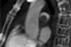
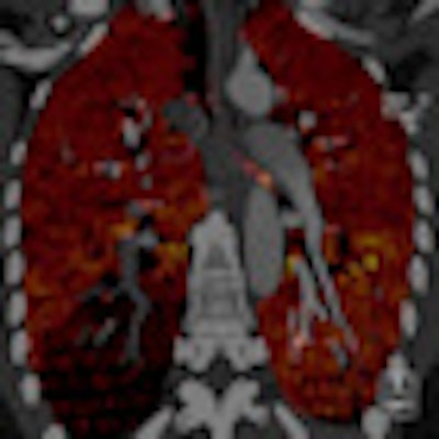
| Above, CTA showing an axial scan with an embolus at the right lower lobe. Below, corresponding coronal pulmonary blood volume map shows a perfusion defect of the right lower lobe. Images courtesy of Tobias de Zordo, MD, and Gudrun Feuchtner, MD. |
 |
Study limitations included a small population that permitted only limited randomization by sex and age. In addition, only three protocols were analyzed, de Zordo said.
On DSCT images, seven patients with pulmonary emboli showed perfusion defects in the corresponding lung parenchyma. In one patient, a triangular perfusion defect was found without a corresponding pulmonary embolus. "We didn't have any additional perfusion defects; we don't know if it's artifact or something else -- the lung window couldn't find anything," he said.
High-pitch dual-source CT allows for significantly reduced radiation exposure, while providing complementary evaluation of perfusion defects, de Zordo concluded.
By Eric Barnes
AuntMinnie.com staff writer
February 18, 2011