Dear CT Insider,
Neurovascular imaging is heading in new directions with the development of 320-detector-row CT scanning. The modality's wide anatomic coverage permits scanning of the entire brain in a single gantry rotation, opening the door to new diagnostic and prognostic possibilities.
For example, radiologists at Nevada Imaging Center in Las Vegas are using 320-row CT to acquire single-volume head scans with time-resolved 4D CT angiography and perfusion images in less than a minute. And they're using the images not only to diagnose disease, but to predict and evaluate treatment response.
For details, images, and videos of their impressive early results, just click on this issue's Insider Exclusive, delivered to our subscribers before other AuntMinnie.com members can access it.
Another head CT story examines soldiers who have suffered traumatic head injuries in the Iraq war. Staff writer Wayne Forrest reports on the Balad Air Force medical center in Iraq, where there is CT but no MRI, and far too many victims of improvised explosive devices. Learn how they're coping by clicking here.
In cardiac imaging, Pennsylvania researchers triaged a fairly sizeable group of chest pain patients successfully and cheaply with coronary CT angiography (CTA). You'll find our exclusive report from the American College of Radiology Imaging Network's (ACRIN) fall meeting here. Meanwhile, 320-slice CTA is scanning the heart in a single rotation, and dual-source CT angiography has hopped onto the ultra-low-dose bandwagon. Dual-source CT is also helping to distinguish malignant lung nodules.
Unfortunately, obese patients are still hard-pressed to find scanning facilities able to accommodate them. Scroll down for links to these and other stories in your CT Digital Community.


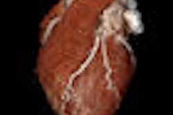


![Axial images from unenhanced calcium score cardiac CT (left) and curved planar reformation images from CT angiography (right) show that higher long-term exposure to air pollution is associated with greater coronary artery calcium and more obstructive coronary artery disease (CAD). Top row: Images in a 68-year-old male patient with higher 10-year mean ambient air pollution exposure (7.9 μg/m3 for particulate matter measuring ≤2.5 μm in diameter [PM2.5] and 17.4 parts per billion [ppb] for NO2) with extensive CAD (coronary artery calcium score [CACS] >1,000 and obstructive CAD [≥70% diameter stenosis]). Bottom row: Images in a 57-year-old female patient with lower 10-year mean ambient air pollution exposure (6.3 μg/m3 for PM2.5 and 4.6 ppb for NO2) with no CAD (CACS = 0 and no obstructive stenosis).](https://img.auntminnie.com/mindful/smg/workspaces/default/uploads/2026/06/hanneman.r6SMLzkezo.png?auto=format%2Ccompress&dpr=2&fit=crop&h=167&q=70&w=250)


