Dear CT Insider,
Pending the results of large, randomized trials, the jury is, of course, still out on the question of whether CT lung cancer screening would reduce mortality and morbidity population-wide. Studies have conflicted famously on the issue, while a more recent report from Massachusetts General Hospital in Boston found that modest mortality gains would result from the routine screening of smokers.
On the other hand, if your early-stage lung cancer is detected and resected successfully, you will almost certainly live longer.
It's also clear that CT needs help to improve the odds. On one level this can mean diligence on the part of radiologists to discern suspicious nodules. Regarding this topic, Dr. Charles White from the University of Maryland in College Park surveys the pitfalls that can lead to missed lung cancers.
On another level it means pairing CT with other promising modalities such as PET. Finally, optimizing CT screening means getting the most out of the image data that's already there, through computer-aided detection (CAD).
Researchers from Catholic University of America and Georgetown University Medical Center in Washington, DC, found surprisingly good results in a pilot study using discrete wavelet transform analysis, a method rarely applied to CAD algorithms.
The researchers compared their method to a laundry list of classical CAD feature parameters to determine which method found the most lung cancers. Their intriguing results are the subject of this issue's Insider Exclusive, brought to you before it's made available to other AuntMinnie.com members.
For another laundry list, including a report on crack lung, and a careful comparison of fourth-generation CT scanners, be sure to scroll down through all of the stories in your CT Digital Community.


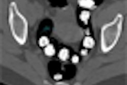
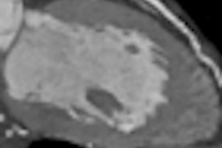


![Axial images from unenhanced calcium score cardiac CT (left) and curved planar reformation images from CT angiography (right) show that higher long-term exposure to air pollution is associated with greater coronary artery calcium and more obstructive coronary artery disease (CAD). Top row: Images in a 68-year-old male patient with higher 10-year mean ambient air pollution exposure (7.9 μg/m3 for particulate matter measuring ≤2.5 μm in diameter [PM2.5] and 17.4 parts per billion [ppb] for NO2) with extensive CAD (coronary artery calcium score [CACS] >1,000 and obstructive CAD [≥70% diameter stenosis]). Bottom row: Images in a 57-year-old female patient with lower 10-year mean ambient air pollution exposure (6.3 μg/m3 for PM2.5 and 4.6 ppb for NO2) with no CAD (CACS = 0 and no obstructive stenosis).](https://img.auntminnie.com/mindful/smg/workspaces/default/uploads/2026/06/hanneman.r6SMLzkezo.png?auto=format%2Ccompress&dpr=2&fit=crop&h=167&q=70&w=250)


