NEW ORLEANS - Performing cardiac CT on a 64-slice scanner, even with a limited field-of-view, can often reveal significant findings outside the heart, which clinicians need to follow up, according to a presentation this week at the American College of Cardiology (ACC) meeting.
"These findings emphasize the need for cardiologists who interpret these studies to be aware of noncardiac findings that may influence the overall multidetector computed tomography results," said Dr. Srikanth Sola, an associate staff physician at the Cleveland Clinic Foundation in Ohio.
In this retrospective study, Sola and colleagues reviewed more than 1,000 multidetector-row CT (MDCT) exams performed on one of two 64-slice scanners (Sensation, Siemens Medical Solutions, Malvern, PA; Brilliance, Philips Medical Systems, Andover, MA) during a one-month period. The researchers evaluated patients for suspected coronary artery disease (n = 443) or assessed patients' pulmonary venous anatomy for pulmonary vein isolation for atrial fibrillation (n = 612). The field-of-view was limited to the area surrounding the heart.
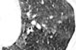
"Of 1,055 patients, 256 had noncardiac abnormalities," said Sola, adding that 10% of these findings were considered clinically significant abnormalities that required additional imaging or clinical follow-up. The significant noncardiac findings included noncalcified lung nodules (> 6 mm), moderate to severe emphysema, pulmonary emboli, and lung cancer.
The results of this study emphasized the importance of cardiologists taking noncardiac findings seriously and sending the patient on for further investigation, commented Dr. John Reilly, the director of cardiac CT at the Ochsner Clinic in New Orleans.
By Ed Susman
AuntMinnie.com contributing writer
March 29, 2007
Related Reading
Radiation dose slashed in 64-slice coronary CTA, February 15, 2007
64-slice CT takes on dual-source in cardiac radiation dose, January 26, 2007
64-slice CT takes on dual-source in cardiac radiation dose, January 26, 2007
Copyright © 2007 AuntMinnie.com