Dear AuntMinnie Member,
The use of CT has grown sharply in the urgent care setting due to its ability to quickly and accurately assess neurological trauma. But there's still debate over exactly when and where to scan, especially when economic and medicolegal considerations come into play.
The current thinking on the use of CT in neurological trauma was the subject of a presentation at this week's Society of Critical Care Medicine meeting in San Francisco. Staff writer Eric Barnes was on the scene to report on the topic for our CT Digital Community.
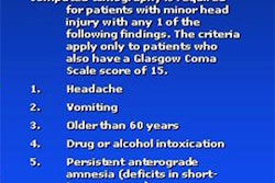
In general, the use of CT for head trauma has been steadily expanding. Indeed, most of the guidelines for assessing neurological trauma tend to encourage it. Trauma physicians have found that the drawbacks of missing pathology by not ordering a CT scan far outweigh the cost of negative scans that might result from a more aggressive posture.
In some cases the need for CT is clear -- C-spine trauma is one such example. But there are still instances when imaging can be skipped entirely. Find out what they are by clicking here.
Also featured this week in the community, Irish researchers cast doubt on the effectiveness of low-dose CT as a screening modality for lung cancer, in a story available here. New results from the International Early Lung Cancer Action Project suggest that patients with secondary lung tumors should be managed more aggressively -- click here for that story. Finally, click here for an article on an effort by German researchers to use CT to differentiate types of noncalcified plaque.
You'll find all these articles and more in our CT Digital Community, at ct.auntminnie.com.


![Axial images from unenhanced calcium score cardiac CT (left) and curved planar reformation images from CT angiography (right) show that higher long-term exposure to air pollution is associated with greater coronary artery calcium and more obstructive coronary artery disease (CAD). Top row: Images in a 68-year-old male patient with higher 10-year mean ambient air pollution exposure (7.9 μg/m3 for particulate matter measuring ≤2.5 μm in diameter [PM2.5] and 17.4 parts per billion [ppb] for NO2) with extensive CAD (coronary artery calcium score [CACS] >1,000 and obstructive CAD [≥70% diameter stenosis]). Bottom row: Images in a 57-year-old female patient with lower 10-year mean ambient air pollution exposure (6.3 μg/m3 for PM2.5 and 4.6 ppb for NO2) with no CAD (CACS = 0 and no obstructive stenosis).](https://img.auntminnie.com/mindful/smg/workspaces/default/uploads/2026/06/hanneman.r6SMLzkezo.png?auto=format%2Ccompress&dpr=2&fit=crop&h=167&q=70&w=250)
