Siemens Medical Solutions reported this week that it has installed its first Somatom Sensation 40-slice CT scanner at the radiology department of Alamance Regional Medical Center located in Burlington, NC.
The new system replaces the 238-bed private, not-for-profit hospital's Somatom Sensation 10 CT unit, according to the Malvern, PA-based Siemens. Experts from the facility will participate in a final round of assessment and feedback for Siemens before the launch of the new 40-slice system.
Worldwide shipment of the Somatom Sensation 40 will start in July this year, Siemens said.
By AuntMinnie.com staff writers
May 17, 2005
Related Reading
Siemens starts Sonoline G40 shipments, May 10, 2005
Siemens debuts magnetic navigation system, May 3, 2005
Siemens, MGH partner on flat-panel volumetric CT prototype, April 28, 2005
Siemens nets HealthTrust contracts, April 22, 2005
Siemens releases Trio with Tim, April 18, 2005
Copyright © 2005 AuntMinnie.com


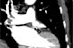


![Axial images from unenhanced calcium score cardiac CT (left) and curved planar reformation images from CT angiography (right) show that higher long-term exposure to air pollution is associated with greater coronary artery calcium and more obstructive coronary artery disease (CAD). Top row: Images in a 68-year-old male patient with higher 10-year mean ambient air pollution exposure (7.9 μg/m3 for particulate matter measuring ≤2.5 μm in diameter [PM2.5] and 17.4 parts per billion [ppb] for NO2) with extensive CAD (coronary artery calcium score [CACS] >1,000 and obstructive CAD [≥70% diameter stenosis]). Bottom row: Images in a 57-year-old female patient with lower 10-year mean ambient air pollution exposure (6.3 μg/m3 for PM2.5 and 4.6 ppb for NO2) with no CAD (CACS = 0 and no obstructive stenosis).](https://img.auntminnie.com/mindful/smg/workspaces/default/uploads/2026/06/hanneman.r6SMLzkezo.png?auto=format%2Ccompress&dpr=2&fit=crop&h=167&q=70&w=250)


