Dear AuntMinnie Member,
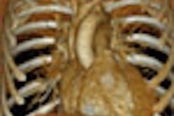
German researchers have taken a technique commonly used in cardiac MRI and adapted it to 16-slice CT imaging of acute myocardial infarction. The protocol takes advantage of late-enhancement patterns in contrast cardiac CT studies to produce images that have good agreement with cardiac MRI scans, and can be used to assess myocardial viability, according to the research team.
Contrast-enhanced CT is a common method of imaging the coronary arteries, but radiologists have tended to focus on what happens to the contrast media during the arterial phase rather than afterward. The group, from the University of Aachen, decided to investigate the intriguing contrast-enhancement patterns that occur in the heart some 15 minutes after the arterial phase.
During this late-enhancement period, areas of infarcted tissue initially appear dark after contrast administration, but brighten over time. The researchers compared these images with both arterial-phase CT and late-enhancement MRI, which is already being used for myocardial viability assessment.
The results were promising, with late-enhancement CT correlating well with delayed-enhancement MRI. Arterial-phase CT images, on the other hand, showed less agreement with either technique.
The encouraging results, along with a handful of other recent studies on the topic, suggest that CT may be ready to add myocardial tissue viability assessment to its capabilities, which already include imaging of the coronary arteries and heart function. Learn more about the group's work by clicking here, and check out other AuntMinnieTV segments by clicking on the links below.


![Axial images from unenhanced calcium score cardiac CT (left) and curved planar reformation images from CT angiography (right) show that higher long-term exposure to air pollution is associated with greater coronary artery calcium and more obstructive coronary artery disease (CAD). Top row: Images in a 68-year-old male patient with higher 10-year mean ambient air pollution exposure (7.9 μg/m3 for particulate matter measuring ≤2.5 μm in diameter [PM2.5] and 17.4 parts per billion [ppb] for NO2) with extensive CAD (coronary artery calcium score [CACS] >1,000 and obstructive CAD [≥70% diameter stenosis]). Bottom row: Images in a 57-year-old female patient with lower 10-year mean ambient air pollution exposure (6.3 μg/m3 for PM2.5 and 4.6 ppb for NO2) with no CAD (CACS = 0 and no obstructive stenosis).](https://img.auntminnie.com/mindful/smg/workspaces/default/uploads/2026/06/hanneman.r6SMLzkezo.png?auto=format%2Ccompress&fit=crop&h=100&q=70&w=100)


![Axial images from unenhanced calcium score cardiac CT (left) and curved planar reformation images from CT angiography (right) show that higher long-term exposure to air pollution is associated with greater coronary artery calcium and more obstructive coronary artery disease (CAD). Top row: Images in a 68-year-old male patient with higher 10-year mean ambient air pollution exposure (7.9 μg/m3 for particulate matter measuring ≤2.5 μm in diameter [PM2.5] and 17.4 parts per billion [ppb] for NO2) with extensive CAD (coronary artery calcium score [CACS] >1,000 and obstructive CAD [≥70% diameter stenosis]). Bottom row: Images in a 57-year-old female patient with lower 10-year mean ambient air pollution exposure (6.3 μg/m3 for PM2.5 and 4.6 ppb for NO2) with no CAD (CACS = 0 and no obstructive stenosis).](https://img.auntminnie.com/mindful/smg/workspaces/default/uploads/2026/06/hanneman.r6SMLzkezo.png?auto=format%2Ccompress&fit=crop&h=112&q=70&w=112)


