A negative or positive 18F-FDG-PET scan can predict Hodgkin’s disease relapse during the post-treatment period, according to Canadian investigators. In addition, PET imaging may be a better option than CT follow-up in these patients.
CT lacks the ability to distinguish viable tumor tissue from fibrosis, whereas previous studies have shown that FDG-PET is an effective tool for staging and monitoring post-therapeutic responses in lymphoma, according to Dr. Christian Guay and colleagues in the Journal of Nuclear Medicine. Guay is from the Université de Sherbrooke in Quebec. His co-authors are from the same institution and from the Centre Hospitalier Universitaire de Sherbrooke.
"Our objectives were to determine the value of 18F-FDG-PET in predicting relapse in relation with the time intervals between the end of chemotherapy and the PET study," they wrote (JNM, August 2003, Vol. 44:8, pp. 1225-1231).
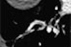
Forty-eight patients with biopsy-proven Hodgkin’s disease were included in this retrospective review. They underwent PET scans on an ECAT EXACT HR+ (Siemens Medical Solutions, Erlangen, Germany), and 2-D images were acquired from the neck to the upper part of the thighs, one hour after IV administration of 7.5 MBq/kg of 18F-FDG. The acquisition times were 5-7 minutes for the emission scan and 3 minutes for the transmission scan for each bed position, covering 5-7 bed positions from the neck to the upper femurs.
"All foci of elevated 18F-FDG uptake not explainable by physiologic uptake were considered to represent viable lymphoma," they said. "The standardized uptake value (SUV) was measured at the most active lesion...a region of interest (ROI) corresponded to 75% of the maximal lesion activity."
CT scans of the pelvis, abdomen, or thorax were done on a Somatom Plus 4 (Siemens) in 32 patients. For thoracic studies, 8-mm slice intervals were obtained; 5 mm for abdominal and pelvic studies. Any lymph node larger than 1 cm in diameter was considered positive for residual lymphoma.
Among the 48 post-therapy patients who underwent 18F-FDG-PET studies, 25% of the scans (12 patients) were deemed positive. Of those dozen patients, 92% (11) eventually relapsed on follow-up. The SUVs of the most active lesion in patients with positive scans ranged from 1.78 to 14.09. The median disease-free interval was 79 days.
 |
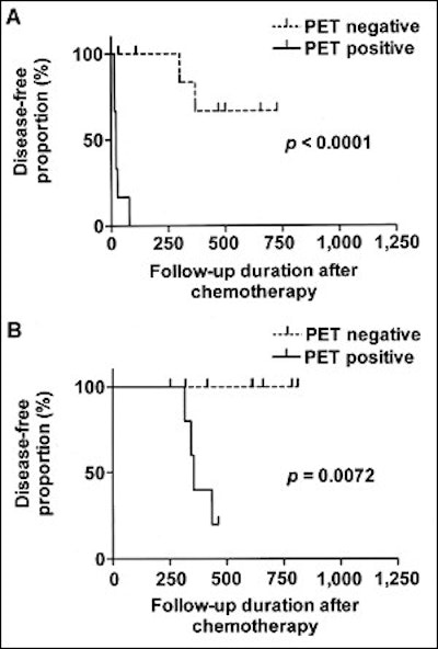
| Kaplan-Meir estimate of disease-free interval. 18F-FDG-PET was performed in the first 30 days after chemotherapy (A) and 91-365 days after chemotherapy (B). Reprinted by permission of the Society of Nuclear Medicine from: Guay, C., et al. Prognostic Value of PET Using 18F-FDG in Hodgkin’s Disease for Posttreatment. J Nucl Med 2003, 44:1225-1231. |
Among the 26 patients with negative results on PET, 8% relapsed on follow-up. The median disease-free survival for these patients was 1,861 days. There were three false-negative PET studies in patients who underwent the scan no more than 49 days after the end of chemotherapy. The authors chalked these results up to the scans being performed too soon.
"No imaging modality can detect microscopic residual disease, and this might be the primary explanation for the negative PET studies, given that the time lag between the negative PET scans and relapse was close to one year in all cases," they said.
Overall, in the 48 patients with positive results, the sensitivity of PET was 79%, the specificity was 97%, and the accuracy was 92%. In comparison, in the subset of 32 patients who also underwent CT imaging after treatment, the sensitivity was 83%, the specificity was 40%, and the accuracy was 56%.
The group concluded that PET was superior to CT for assessing residual disease after therapy. However, they acknowledged a potential bias in the study in that not all of the patients underwent both PET and CT imaging, which would be closer to a real-life clinical scenario.
"Oncologists rarely rely on a single modality in the decision-making process, and an integration of several data sources with confirmatory studies is generally used to guide therapy," they wrote. "A potential research avenue that remains to be explored is determination of the (value) of 18F-FDG-PET as a prognostic indicator very early after initiation of treatment."
By Shalmali PalAuntMinnie.com staff writer
September 1, 2003
Related Reading
Risk of breast cancer after treatment for Hodgkin disease quantified, July 23, 2003
Chemotherapy reduces breast cancer risk in radiation-treated Hodgkin's patients, July 2, 2003
Radiation for Hodgkin lymphoma before age 30 ups breast cancer risk: study, June 30, 2003
Focal liver lesions detected during NHL follow-up need full diagnostic work-up, January 24, 2003
MRI shows potential for staging pediatric non-Hodgkins lymphoma, January 2, 2003
Copyright © 2003 AuntMinnie.com