Japanese researchers are relying on high-resolution CT to spot increasingly smaller lung lesions, according to a presentation at the 2002 Society of Thoracic Surgeons meeting in San Diego earlier this month. On the CT scan, these ground-glass-opacity lesions have a far different prognosis than do similar-sized solid lesions.
"These ground-glass-opacity lesions are true early cancers," said Dr. Hisao Asamura, a researcher at the National Cancer Center Hospital in Tokyo. His CT studies indicate that the ground-glass-opacity lesions progress to becoming partially solid and then solid.
The ground-glass lesions -- which are clear enough for visualization of blood vessels that are underneath the lesion -- appear to have few invasive characteristics, he explained. On the other hand, solid tumors, even those that are less than a centimeter in size, have invasive characteristics and require lobectomy, he said. But in some cases the treatment of smaller, ground-glass lesions can be curative without major lung resection, he said.
In his study, Asamura reviewed findings of lung tumors less than a centimeter in diameter that were found in CT screening programs, or were found incidentally through x-ray and CT procedures for other indications.
In a review of data collected over the past 10 years, Asamura and colleagues noted that 1,769 lung cancers were resected. Of that group, 48 were less than a centimeter in size and fell into three categories:
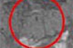
- Simple ground-glass-opacity type. There were 19 of these tumors, which did not obscure the bronchovascular structure. The histology showed these tumors were bronchioloalveolar carcinoma or adenocarcinoma with predominantly bronchioloalveolar spread.
- Complex ground-glass opacity. Seen on 9 CT exams. These lesions contained a solid area as well as a clear area. The histology of these tumors was similar to the simple ground-glass-opacity lesions.
- Solid lesions. The researchers identified 20 of these lesions. These tumors were also predominantly adenocarcinomas, but two were squamous cancer, one was small-cell lung cancer and was one characinoid. Lymph-node involvement was observed only in three solid lesions.
Limited resection was performed in 15 of the 29 tumors showing ground-glass expression, and in four of the solid tumors. Tumor recurrence occurred in two solid lesions; one each in bone and locoregional lymph nodes. There were no cancer-related deaths.
For all three lesion types, age and gender distributions were similar, with an average of 61 years and almost even male-to-female distribution. Twenty-six tumors were screen-detected; the others were found incidentally.
By Edward SusmanAuntMinnie.com contributing writer
February 10, 2003
Related Reading
Lung cancer screening not cost-effective, say Johns Hopkins researchers, January 14, 2003
R2 and Vital Images to merge CT lung nodule software, December 4, 2002
Sputum cytology gives a big boost to CT lung screening, December 2, 2002
CAD highlights hazy opacities on chest x-rays, October 11, 2002
Copyright © 2003 AuntMinnie.com