BOSTON - The future of cardiovascular disease assessment will be multidisciplinary. Laboratories will measure C-reactive protein and perform advanced lipid analysis. They'll look for homocysteine, cell adhesion, and genetic markers to evaluate the physiological culprits underlying subclinical heart disease. In imaging, ultrasound and nuclear medicine have established their value for certain applications, and nothing depicts the soft tissue of vessel walls like MRI.
But CT will remain an important tool for cardiac assessment, according to several experts who spoke at this week's Society of Computed Body Tomography and Magnetic Resonance’s course on medically principled CT screening.
Over the next 5-10 years, researchers will work to define the best ways to combine the technologies to identify at-risk individuals and intervene to keep them healthy, according to Dr. Jeffrey Carr, from the departments of radiology and public health at Wake Forest University in Winston-Salem, NC.
One of his talks on Saturday focused on CT cardiac assessment and calcium scoring, on technical improvements in the modality, and on a new population-based trial that promises to help determine how to fit the new technologies together.
"We've known about coronary calcium from the very beginning, so a big problem is individuals not learning from the past," he said. "There is persuasive evidence that understanding calcified plaque is a big part of understanding who's going to go on to have coronary events."
So-called hardening of the arteries has been associated with cardiovascular disease for nearly 50 years, but a 1980 study suggested a strong relationship. Margolis and colleagues found a significant difference in five-year survival between those with coronary calcium (58%) and those without it (87%). The findings were independent of gender, the number of diseased vessels, exercise test results, or left ventricular function (Radiology, December 1980, Vol. 137:3, pp. 609-616).
Then, in a 1994 study, Detrano and colleagues prospectively examined 1,400 individuals at high risk for cardiovascular disease. Individuals with coronary calcium at fluoroscopy were three times as likely to experience revascularization, new-onset angina, myocardial infarction, or sudden death. (Journal of the American College of Cardiology, August 1994, Vol. 24:2, pp. 354-358).
Dr. Joseph Shemesh and colleagues followed up in the mid-1990s with studies that elegantly established the heightened risk of cardiovascular events in several different population groups, Carr said.
There is evidence that the calcified vessels themselves are less likely to rupture than other vessels in the same patient containing plaque that is more vulnerable. However, the relationship between the patient who has calcium and his or her increased risk of adverse events has held fast. Much effort will be needed over the next several years to determine how calcium scores fit into other risk markers, what the cutoff points are, how scoring can be improved, and whether any meaning can be extracted from changes in baseline total calcium score, he said.
Instrumentation advances
A major step forward in cardiac CT imaging was the development of the electron beam CT scanner in the early 1980s, Carr said. The dual-slice helical scanner (CT Twin, Elscint) became available in 1995; subsecond helical CT with ECG gating in 1999. In 2000, the introduction of 4-detector multidetector-row scanners improved temporal resolution to 300 msec.
In 2001, the introduction of sector, or "burst" mode, improved temporal resolution to 130 msec. This year, the introduction of 16-detector machines again speeded scanning times and improved resolution. In 2003, volume CT acquisition will enable imaging of the heart in a single acquisition with near-isotropic resolution, Carr said.
Carr and colleagues' current cardiac CT protocol uses 50 to 100 mAs, depending on whether it's a standard or low-dose acquisition, 120 kVp, and prospective gating, which means the scan is being acquired in axial or cine mode, the most dose-efficient mode, he said.
"Gantry rotation should be 0.5 seconds or (faster)," Carr said. "It's possible to do cardiac CT with a 0.8-second gantry rotation, but 0.5-second gantry rotation provides a significant improvement in image quality. Our slice collimation is 2.5 mm, and the display field of view can range between 26 and 35 cm. We use what's called a segmented or partial-scan reconstruction," with a 220-degree field of view to improve temporal resolution.
ECG gating is telling the scanner where to acquire the image in the systolic/diastolic waveform, and while there has been debate on this issue, his group has found the best results using an area in mid-diastole.
"One of the advantages of MDCT vs. single-slice CT is the idea of slice registration. If you take a (single) slice at each cardiac cycle there is beat-to-beat variability. So your artery is going to be offset based on this variability. The advantage of MDCT is that you're actually acquiring four levels on a four-channel system simultaneously within the cardiac cycle. These four levels are perfectly aligned, with no gaps and no overlap, so you can reduce the beat-to-beat variability because the slices are perfectly aligned."
A current goal for most manufacturers is volume CT, which will enable acquisition of most of the coronary arteries in one pass, six slices, and a breathhold of just 8-14 seconds for an 8-channel scanner. In comparison, a single-slice scanner would require 48 slices at 2.5-mm collimation to cover 120 mm, and the patient would have to hold his breath for 80-160 seconds.
"Only pearl divers can do this," Carr said.
Calcium scoring
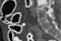
It's important to remember that the standard Agatston calcium scoring system isn't based on a sum, but rather the area of the plaque multiplied by a weighting factor, itself based on the pixel in the plaque volume with the highest CT value not less than 130. As such, it's an arbitrary system that will also yield to refinement as calcium scoring progresses, he said.
"There's nothing in the pathobiology of atherosclerosis that says that calcified plaque at a lower CT number is any different from a higher CT number," Carr said. "We’re truncating what we're able to measure based on the technology we have."
The weighting factor is zero for pixels under 130 heat units, 1 for 130-199, 2 for 200-299 and so forth. It is the weighting factor that changes the difference between volume measurement of the size of the plaque and the density of the plaque, Carr said.
A couple of studies have compared MDCT to EBCT scanners for coronary calcium screening, and the results were very comparable, except in the low scores, which can vary widely, Carr said. But low-score variability is also seen in repeat studies of the same patient on the same scanner.
"We're seeing the physiology of the motion of coronary arteries and the limitations of the technology," he said. "If your slice collimation is 3 mm or 2.5 mm, you have a big effect from partial-voluming. And so a lot of the variability is simply related to the motion of the coronary artery, and plaques falling between the slices."
Coronary calcium screening features prominently in a new study his group is participating in, Carr said. The Multi-Ethnic Study of Atherosclerosis (MESA) will screen some 6,500 men and women -- Asian, black, Hispanic, white -- ages 45-84, who are asymptomatic for cardiovascular disease at baseline.
"We'll do a comprehensive assessment of cardiovascular disease risk, and subclinical coronary disease," he said. "We're doing cardiac MR and cardiac CT, carotid ultrasound, brachial artery ultrasound, extensive lab testing, lipids, inflammatory markers, a large number of questionnaries -- and then we measure the current traditional CVD risk factors of blood pressure, height, weight, etc."
MESA will provide the first true sampling of the distribution of cardiovascular disease and plaque burden across the population, he said. Comprehensive analysis of the data will attempt to validate the spectrum of tests and biological markers being used, including the utility of each test and its effectiveness in relation to other tests.
Another innovation will be the use of a special phantom containing known calcium levels. The phantom will be imaged right along with the patient, he said. That way, the CT images can be adjusted for each dataset, like a test pattern, to provide more accurate calcium scoring. The first year's CT data will be presented at the American Heart Association meeting later this month, Carr said.
"We can measure coronary calcium accurately and reproducibly with EBCT/MDCT," Carr concluded. "The advancing technology is going to make the quantification of calcified plaque easier and better, and we're developing calibration methods that will allow us to further reproduce the measurements of coronary calcium."
By Eric BarnesAuntMinnie.com staff writer
November 13, 2002
Related Reading
Multislice spiral computed tomography improves CAD diagnosis, October 2, 2002
In elderly, coronary calcium is strongest predictor of mortality, March 1, 2002
Copyright © 2002 AuntMinnie.com