Dark-field chest x-ray could be useful for determining optimum mechanical ventilation pressure to use in hospitalized patients, according to researchers at the Technical University of Munich (TUM).
In a cadaver study, the group tested the capabilities of a dark-field chest x-ray prototype for measuring signal changes generated by the lung at different levels of inflation, such as those experienced by hospitalized patients requiring mechanical ventilation. The approach allowed for quantitative analysis of the subject's lung inflation cycle and suggests that the technique could be used to avoid ventilator-induced lung injury, they wrote.
"Particularly in patients with acute respiratory distress syndrome (ARDS), it is important to both improve lung function and decrease the chance of developing ventilator-induced lung injury," wrote lead author Dr. Florian Gassert in a research letter published December 15 in Radiology: Cardiovascular Imaging. "Thus, a marker indicating the condition of lung alveoli at different pressure levels is highly desirable,"
Dark-field radiography is an emerging technique for the imaging of pulmonary diseases. Signal is generated by small-angle scattering of x-rays due to multiple refractions at tissue-air interfaces in lung alveoli. First studies in humans postulate that the quantitative dark-field signal of the lung correlates with the number of tissue-air interfaces in lung tissue, yet the dark-field signal has been analyzed only at full inspiration in humans, the authors noted.
In this study, Gassert and colleagues performed imaging on a 78-year-old man two days after he died from cardiac arrest. His underlying disease was osseous metastatic prostate cancer, with no pulmonary metastasis. The cadaver was intubated and inflated at different pressure levels (at 0.0, 1.0, 2.0, and 3.0 kilopascals [kPa], or 0.0, 0.15, 0.29, and 0.44 pounds per square inch [psi]) in three cycles. For every ventilation pressure and cycle, the researchers calculated total signal of the analyzed area for both dark-field images and conventional images in meters squared.
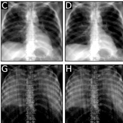
 Conventional (upper row) and dark-field (lower row) radiographs of the first inflation cycle at (A, E) 0.0 kPa (0.0 psi), (B, F) 1.0 kPa (0.15 psi), (C, G) 2.0 kPa (0.29 psi), and (D, H) 3.0 kPa (0.44 psi). For both imaging modalities, greater lung volume can be observed with higher pressure. Attenuation signal of the lung visually decreases with higher pressure levels, while dark-field signal increases. Image courtesy of Radiology: Cardiothoracic Imaging.
With higher pressure, the segmented lung area had higher visual and quantitative signal intensity on both dark-field and attenuation images, the investigators found. Overall, dark-field signal intensities at the same pressure were higher at later cycles.
Specifically, dark-field signal intensity was lower when pressure changed from 0.0 kPa to 1.0 kPa (0.00 psi to 0.15 psi) and thereafter, it followed the same pattern as that of the entire lung. Moreover, the attenuation signal behaved opposite to the dark-field signal, with a lower signal at higher pressures and later cycles, the researchers found.
"Dark-field radiography in a cadaveric lung showed signal changes in structurally unimpaired alveoli following mechanical inflation at different pressures, demonstrating that the dark-field signal is generated by tissue-air interfaces," they wrote.
While the study was performed in a single human cadaver, it may be an important step toward the analysis of dark-field signals in living humans at different inspiration levels, the group concluded.