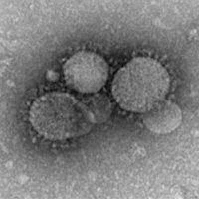
The first case of Middle East respiratory syndrome (MERS) coronavirus infection has appeared in the U.S., reported U.S. government officials on May 2.
The case was confirmed in a traveler from Saudi Arabia who has been hospitalized in Indiana, according to officials with the U.S. Centers for Disease Control and Prevention (CDC). The virus was first reported in Saudi Arabia in 2012; recent scientific reports have linked the virus to camels.
The traveler left Saudi Arabia on April 24, traveling by plane from Riyadh to London and then Chicago, where the individual took a bus to Indiana. On April 27, the patient began to experience symptoms including shortness of breath, coughing, and fever, and was admitted to a hospital on April 28.
Indiana public health officials tested for MERS infection due to the patient's travel history, and they confirmed the case on May 2. The patient is in stable condition in isolation and is being well-cared for, according to the CDC.
The case presents a very low risk to the general public, according to Dr. Anne Schuchat, assistant surgeon general and director of the CDC's National Center for Immunization and Respiratory Diseases. While in some countries MERS has spread through close contact, such as caring for or living with someone who is infected, there is no evidence of infection through community settings.
Health officials are not sure how the patient was infected with MERS; exposure to the virus may have taken place in Saudi Arabia, but it's unknown how many people had close contact with the patient.
Including the Indiana case, there have been 401 confirmed cases of MERS infection in 12 countries, with all reported cases originating in six nations in the Arabian Peninsula. Most individuals with MERS infection have developed severe acute respiratory illness, and 93 people have died, the CDC said.





![Examples of ultrasound findings and techniques. (A) Images in a 39-year-old male patient with a mass in the left thigh. The mass is heterogeneous on the B-mode US image (compared with the patient in D) and showed increased microvascularity (superb microvascular imaging [SMI]) and shear-wave elastography (SWE) values. Undifferentiated pleomorphic sarcoma was diagnosed at biopsy (with pleomorphic rhabdomyosarcoma in surgical specimen). (B) Images in an 18-year-old male patient with a mass in the left leg. The mass is hypoechoic on the B-mode image, with no other findings suggestive of malignancy. The lesion is in contact with the cortex of the tibia, which is slightly irregular. CT revealed a doubtful anteromedial tibial erosion. The microvascular study demonstrated high vascularization, suggestive of malignancy. Periosteal Ewing sarcoma was diagnosed with both histologic and immunohistochemical confirmation. (C) Images in a 69-year-old female patient with a lump growing on the outside of the left leg. Multiple SWE examinations were performed (please note the high values obtained in the measurements, whereas the color map highlights the stiffness relative to adjacent tissues). SMI showed areas of increased vascularization to target for sampling. Undifferentiated spindle cell sarcoma was diagnosed at biopsy, with residual leiomyosarcoma in the surgical specimen after neoadjuvant therapy. (D) Images in a 56-year-old female patient with a mass in the right thigh. The mass is heterogeneous at both B-mode ultrasound (similar to patient A) and MRI (coronal T2-weighted spectral attenuated inversion recovery [SPAIR]; T1-weighted pre-contrast and postcontrast imaging), which even shows uptake after the administration of paramagnetic contrast material, which is traditionally suggestive of malignancy. Low values at SMI and elastography are suggestive of benignity. Spindle cell lipoma was diagnosed at biopsy, with atypical spindle cell lipomatous tumor in the surgical specimen.](https://img.auntminnie.com/mindful/smg/workspaces/default/uploads/2026/08/images-radiol250278fig2.APCFLSvX6p.jpg?auto=format%2Ccompress&fit=crop&h=112&q=70&w=112)


