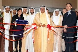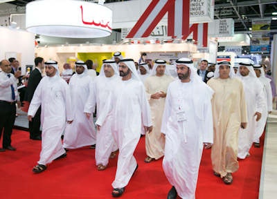
DUBAI - The ruler of Dubai, Vice President, and Prime Minister of the United Arab Emirates Sheikh Mohammed bin Rashid Al Maktoum, visited the Arab Health meeting on Tuesday. Accompanied by Dubai Crown Prince Sheikh Hamdan bin Mohammed bin Rashid Al Maktoum and Dubai Deputy Ruler Sheikh Maktoum bin Mohammed bin Rashid Al Maktoum, Sheikh Mohammed toured the exhibition. His group visited the GE Healthcare and the Cleveland Clinic booths.
According to Sheikh Mohammed's official website, he expressed happiness over the United Arab Emirates' hosting of such prestigious exhibitions and world gatherings which, he said, contribute to the national economy and form a platform for exchange of expertise and knowledge.
Also accompanying Sheikh Mohammed on his tour were Chairman of Dubai Civil Aviation and Chief Executive of Emirates Group Sheikh Ahmed bin Saeed Al Maktoum, Acting Health Minister Abdul Rahman Mohammed Al Owais, Dubai Health Authority Director General Eng. Eisa Al Maidoor, Dubai Director of Protocol and Hospitality Khalifa Saeed Suleiman, CEO of Dubai World trade Center Hilal Saeed Al Marri, and other senior officials.
 The ruler of Dubai, Sheikh Mohammed bin Rashid Al Maktoum, visited the Arab Health meeting on Tuesday. Accompanied by Dubai Crown Prince Sheikh Hamdan bin Mohammed bin Rashid Al Maktoum and Dubai Deputy Ruler Sheikh Maktoum bin Mohammed bin Rashid Al Maktoum, Sheikh Mohammed toured the exhibition. Photo courtesy of Informa Exhibitions.