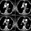
Type A (DeBakey I) Aortic Dissection:
(Click the small images to view the larger radiographs)
The elderly female shown in the case below presented with right flank pain. A non-contrast CT scan of the abdomen was performed which demonstrated displaced intimal calcification (arrow on coned view) within the abdominal aorta.
Non-contrast CT: 

A contrast enhanced CT scan was then performed which demonstrated a Type A aortic dissection that severely compromised the true lumen of the ascending aorta. There was no perfusion to the right kidney which was being supplied via the false lumen:

An angiogram demonstrated the point of rupture within the ascending aorta (arrow). A chest radiograph was obtained prior to the angiogram which demonstrated enlargement of the thoracic aorta (click here to view the CXR):


