Reactivation tuberculosis:
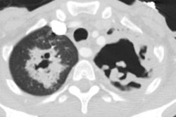
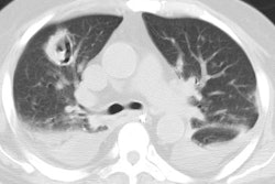
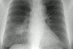
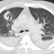
The patient show in the radiographs below presented with a cough. The chest radiograph demonstrated a cavitary infiltrate in the left upper lobe and a separate infiltrate in the left apex. There was no evidence of hilar adenopathy or pleural effusion. The patient had a positive PPD and positive sputum culture for Myobacterium tubersulosis.
(Click on small images to view larger radiographs)
Chest X-ray:
Coned View: (Yellow arrow points to cavitary infiltrate, red arrow to apical infiltrate)

