Case 1: Lymphangioleiomyomatosis
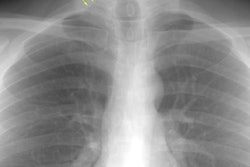
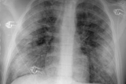
The patient shown below presented with chest pain and shortness of breath. The CXR revealed coarsened, prominent lung markings with a predominantly subpleural right pneumothorax. (Click image to enlarge)

CT was performed and demonstrated multiple cysts throughout the lungs. The pneumothorax was noted to be loculated and predominantly in the lower portion of the right hemithorax.


Fat containing lesions were noted in both kidneys consistent with angiomyolipomas. Subsequently, it was learned that the patient had a history of lymphangioleiomyomatosis and had a prior pleurodesis.