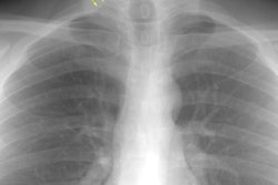
Acute eosinophilic pneumonia:
The patient shown below presented with respiratory distress. The chest radiograph demonstrated bilateral diffuse, but predominantly peripheral areas of airspace consolidation. The bronchoalveolar lavage fluid demonstrated a 37% eosinophil count. The patient demonstrated rapid clinical and radiographic improvement with steroid therapy. (Click on image to enlarge)