Wegener's granulomatosis Overlap syndrome
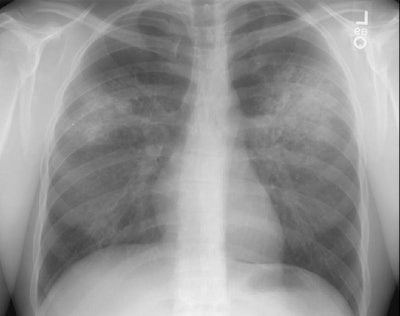
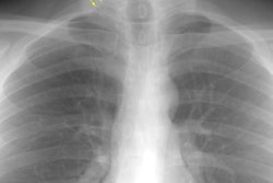
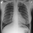
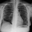
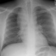
The patient shown in the image below had an extensive travel history and his parents were missionaries in New Guinea. He was referred to the rheumatology service for complaints of polyarthralgia. On exam, additional history revealed complaints of epistaxis, and later hemoptysis. A chest radiograph was obtained.
The chest radiograph demonstrated bilateral consolidations which were primarily confined to the superior segments of the lower lobes and posterior upper lobe segments (click here to see the lateral exam). A PPD was placed and was negative, and the patient was HIV negative. Although plain films of the paranasal sinuses were normal, on direct visualization, mucosal ulcerations were identified. At bronchoscopy, blood was noted within the airways- indicating that the parenchymal abnormalities were likely the result of hemorrhage. Both serum and BAL fluid were C-ANCA positive and a diagnosis of Wegener's was made. However, the open lung biopsy specimen demonstrated both features of Wegener's and an eosinophoilic vasculitis (Churg-Strauss) and the final diagnosis was that of an overlap vasculitis. The patient was treated with steroids and methotrexate with a rapid clearing of the parenchymal abnormalities.
