Wegener's Granulomatosis
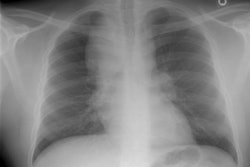
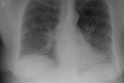
The patient shown in the case below presented with hemoptysis.
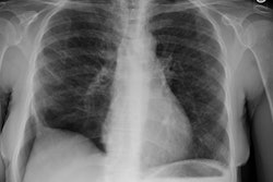
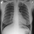
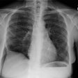
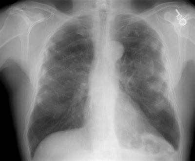
CXR demonstrated cavitary areas of consolidation in the right mid and left upper lungs (Click to enlarge image)
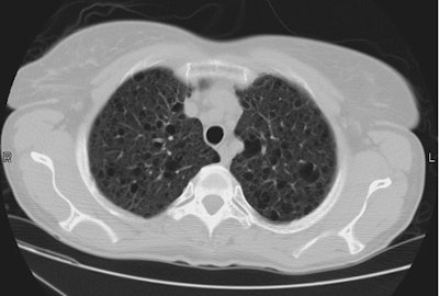
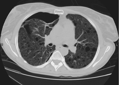
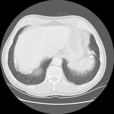
Chest CT revealed cavitary nodules, cavitary areas of consolidation, and non-cavitary areas of patchy parenchymal consolidation. Patient was C-ANCA positive and lung biopsy revealed Wegener's.
