New MRI research suggests the neurological health of boxer athletes must be monitored and maintained long-term, according to research shared May 6 at the International Society for Magnetic Resonance in Medicine (ISMRM) meeting.
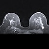
In a poster presentation, researchers from Lanzhou University Second Hospital and GE HealthCare MR Research in China shared results from their analysis of sport-related concussion (SRC) macro- and microstructure alteration patterns in the MRI brain scans of 57 active young boxers with a history of SRC. The study also included 72 healthy exercise-loving controls with non-SRC. The researchers found that the boxers had significant micro- and macrostructural brain changes.
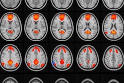
All subjects underwent the same neuropsychological assessment before their MRI scan. The MRI examinations were performed on a 3-tesla MR scanner (SIGNATM Premier) with a 48-channel head coil, according to a poster preview.
Two types of MR imaging, in particular, showed lower white-matter volume and significant white-matter abnormalities possibly associated with SRC in young boxers. SRCs from boxing are common. It is thought that boxers sustain countless concussions and subconcussions during their careers. However, the pattern and extent of damage caused by boxing-related concussions is unknown.
Using 3D T1-weighted imaging and neurite orientation dispersion and density imaging (NODDI), Wenjing Huang and colleagues reported on white matter microstructure, gray- and white-matter volumetric analyses, the cavum septum pellucidum (CSP), and other points of interest from their study.
For example, with NODDI, Huang and colleagues shared that data was obtained with repetition time (TR) = 5,705 ms, echo time (TE) = 68.8 ms, field of view (FOV) = 240 mm, matrix = 120 × 120, 78 slices, slice thickness = 2 mm. Additionally, the NODDI protocol consisted of acquiring diffusion-weighted images along three b-values: twenty directions at b = 1,000 s/mm2, forty directions at b = 1,800 s/mm2, and sixty directions at b = 2,500 s/mm2. Further, NODDI fitting was performed using the CUDA Diffusion Modelling Toolbox (cuDIMOT) running on graphics processing units.
Among the results reported, volumetric analyses revealed the ratio of gray matter volume to total intracranial volume (TIV) and the ratio of cerebral spinal fluid (CSF) volume to TIV increased, and the ratio of white-matter volume to total intracranial volume (TIV) reduced (p < 0.05). The researchers also said that the presence of CSP was 57.1% in the SRC group and 38.9% in the healthy control group (p = 0.04), adding the length of the CSP and the ratio of CSP length to septum length were significantly greater in boxers with SRC. Moreover, the researchers reported their MRI-derived indices of white-matter microstructure.
"For boxers with recurrent chronic SRC, the histopathological features of white matter were characterized by axonal degeneration and demyelination, as well as reversible axonal remyelination and irreversible axonal breaks, which may be associated with diminished [Neurite Density Index] NDI," wrote Huang et al. "Boxers who suffer repeated concussions may involve multifocal damage to myelin proteins in the subcortical white matter, resulting in diffuse changes and reduced white matter volume."
While their report adds to previous studies, the researchers said further studies are needed to investigate the pathologic mechanisms underlying the development of CSP after repetitive head impacts.