Combining MR spectroscopy (MRS) with dynamic, contrasted-enhanced MRI in the breast results in higher sensitivity and specificity, according to Italian radiologists. They presented the results of their study at the 2005 RSNA meeting in Chicago.
"We know that MR for breast has gained a real role in the diagnosis and management of breast cancer.... Our purpose was to test the sensitivity of breast MRS as a final step after dynamic MR imaging and evaluate the prognostic gain using this method," said Dr. Alfonso Fausto from the University Hospital Istituto Policlinico San Donato in Milan.
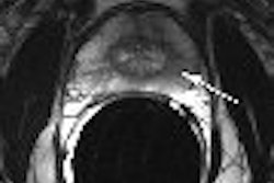
The patient population consisted of 254 patients who underwent T1-weighted, 3D gradient-echo gadolinium-enhanced MRI on a 1.5-tesla scanner. Of these patients, 127 had single-voxel, water- and fat-suppressed spin-echo MRS images obtained, using a PRESS sequence. An intensity choline peak equal to or higher than 2 was considered a sign of malignancy. The gold standard was core biopsy results or one-year follow-up with negative results.
According to the results, 151 voxels with a median size of 2.9 cm3 were obtained on MRS. Three cases had no reliable MR spectra. These cases were ultimately diagnosed as one benign enhancement and two invasive ductal carcinomas. In the remaining spectra, pathology demonstrated 68 malignancies and 80 benign cases. MRS and MRI had seven false negatives combined. Separately, MRS turned in nine false positives while MRI had 15.
Overall, both modalities had a sensitivity of 90%. For MRS the specificity was 89%, the positive predictive value (PPV) was 87%, and the negative predictive value (NPV) was 91%. For MRI the specificity and PPV were both 81% and the NPV was 90%. An 8% gain in specificity was achieved because of the addition of MRS, the authors stated.
"We can see that MRS can be routinely added to the dynamic (MRI) study. We can increase sensitivity if we exclude lesions less than 1 cm. More work must be done to reduce the mean voxel and to adjust for the movement of the patient," Fausto concluded, adding that more work needed to be done to reduce voxel size through sequencing and improve postprocessing.
Fausto and colleagues' work is in line with a previous study by researchers from Johns Hopkins University School of Medicine in Baltimore. This group investigated the feasibility of combined dynamic, contrast-enhanced MRI with MRS in evaluating breast lesions, and suggested that "MRS appears to be a promising technique for classification of breast lesions when (MRI) results are equivocal ... and may have improved specificity compared to either modality alone" (Journal of Magnetic Resonance Imaging, January 2005, Vol. 21:1, pp. 23-28).
By Shalmali Pal
AuntMinnie.com staff writer
December 16, 2005
Related Reading
Breast MRS proves sensitive to all cancers but not in normal breast tissue, November 27, 2005
MRI continues to make headway in breast cancer imaging, October 28, 2005
MR spectroscopy aids breast MR imaging, July 29, 2005
Gadobenate dimeglumine MR spots abnormal breast vascularity, cancer, June 8, 2005
Copyright © 2005 AuntMinnie.com