Thieme, New York, 2002, $129
In major academic centers and in the most advanced private practices, MRI has become the workhorse modality of contemporary neuroradiology. CT has become an easy screening tool for patient triage, but one that ultimately lacks the necessary sensitivity and resolution to serve as an end-stage imaging modality.
Nevertheless, CT is still the main diagnostic tool in neuroradiology, especially among physicians who face geographic and financial constraints. CT of the Head and Spine will be most useful to this group as an overview of the use of CT in diagnosing diseases of the brain and spine.
The book opens with a cursory review of neuroanatomy referenced on axial head CT slices. This is followed by a nice review of the basic clinical and technical aspects of CT, which will be very helpful to the beginner but somewhat mundane to the experienced neuroimager.
The text is divided into sections on traumatic brain injury, vascular disease, inflammatory disease, neoplasm, and demyelination. The facial skeleton and skull base is treated as a single section, followed by spinal anatomy and a rather comprehensive discussion of specific diseases of the spine.
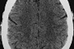
The strengths of this work include numerous small but high-quality images. A thorough review of traumatic brain injury is pertinent to those who rely on the speed and accessibility of CT in emergency rooms as a first-line diagnostic tool for evaluating head trauma. The section dealing with skull base pathology and the facial skeleton is worthwhile, given the value added by CT in many types of maxillofacial disease processes.
One of the main weaknesses of this book is its timeliness -- or lack thereof. The text’s cover depicts several CT images processed from a 3-D workstation. The use of volume rendered images with multiplanar reconstruction software would suggest that advanced CT applications, such as multidetector acquisition and postprocessing techiniques, are reviewed in the text.
Unfortunately, the book contains very little in the way of advanced techniques, and is instead dominated by standard axial CT images and rehashed diagnostic information. Additionally, the labeling of several anatomic images are confusing, with reference lines that are not placed directly on the structure labeled, or structures that are incorrectly labeled altogether. For example, on page 74 there is a mislabeled angiogram supposedly depicting vasospasm at the origin of the middle cerebral artery.
In the end, CT of the Head and Spine has some interesting information, but offers nothing new. For those who must rely on CT as their main neuroimaging modality, this book will be of some value; for those who have both the experience and equipment to conduct more refined CT exams, other comprehensive reference texts would be a better investment.
By Dr. Brian J. FortmanAuntMinnie.com contributing writer
September 5, 2002
Dr. Fortman is a neuroimaging specialist at Carolina Radiology Associates, in Myrtle Beach, SC. He recently completed a neuroradiology fellowship at The Johns Hopkins Hospital in Baltimore.
If you are interested in reviewing a book, let us know at [email protected].
The opinions expressed in this review are those of the author, and do not necessarily reflect the views of AuntMinnie.com.
Copyright © 2002 AuntMinnie.com