In a small series of patients with the swine flu (S-OIV or H1N1) virus, CT was superior to radiography for depicting both lung abnormalities and the distribution of influenza-related disease. Still, radiography detected abnormalities in all cases that were later deemed abnormal on CT, according to an article appearing Wednesday in the American Journal of Roentgenology.
Descriptions of radiologic manifestations of swine flu have been limited to a few case reports, noted Amr Ajlan and colleagues from Vancouver General Hospital.
Most reported findings "were those of unilateral or bilateral; focal, multifocal, or diffuse; ground-glass opacities, consolidation, or interstitial markings," they wrote (AJR online, October 14, 2009). The present study aimed to review the chest radiographic and CT findings in seven patients with confirmed S-OIV infection.
The study group consisted of six men and one woman (mean age, 43 years; range, 16-73 years). Six of seven patients had underlying comorbidities including asthma, multiple myeloma, sickle cell anemia, hypertension, and coronary artery disease.
Chest x-rays were obtained in all seven patients, including two on a mobile unit. Two patients underwent MDCT on admission; a third was scanned within 24 hours of admission, and one received IV contrast because of clinical suspicion of pulmonary embolism.
Three patients had abnormal findings at radiography, including one with faint ground-glass opacities, which progressed from the lower to the middle zone of the right lung. A second patient had extensive bilateral ground-glass opacities and poorly defined areas of consolidation. A third showed a hazy increase in density, the authors wrote.
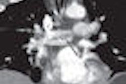
MDCT was initially normal in a patient with lung transplantation, but follow-up MDCT at 12 days showed extensive ground-glass opacities and mild multifocal areas of consolidation. In another patient, initial MDCT showed bilateral multifocal asymmetric ground-glass opacities. A third MDCT case showed bilateral asymmetric ground-glass opacities and areas of consolidation with peribronchovascular predominance.
"In all the patients with abnormal radiographic findings, the MDCT study showed more extensive involvement," they wrote. "In addition, MDCT was superior to radiography in showing the distribution of the disease. Again, ground-glass opacities with multifocal consolidation were the predominant findings in our group of patients."
The researchers said that an interesting observation on the MDCT scans was the distinctive peribronchovascular (n = 3) and peripheral (n = 2) distribution of the disease, which was similar in appearance to cases of organizing pneumonia.
Related Reading
CT reveals acute PE risk in patients with severe swine flu, October 21, 2009
CT can aid early swine flu diagnosis, October 14, 2009
Virus outbreaks put scrutiny on infection control practices, August 10, 2009
Pulmonary CTA finds more than pulmonary embolism in children, August 25, 2009
CT protocols reduce risk for pulmonary embolism, March 9, 2009
Copyright © 2009 AuntMinnie.com