An automated whole-breast ultrasound system doubled the cancer detection rate when used with mammography to screen a population of women with dense breasts and/or an elevated breast cancer risk, according to a multicenter study published online this month in European Radiology.
In addition to doubling the detection rate for all cancers, the system tripled the detection rate for very small cancers -- those smaller than 10 mm. The authors believe the technology's effectiveness could justify adding it to the screening routine for some women.
"Additional detection and the smaller size of invasive cancers may justify this technology's expense for women with dense breasts and/or at high risk for breast cancer," wrote a research team led by Dr. Kevin Kelly of Huntington Memorial Hospital in Pasadena, CA (European Radiology, September 3, 2009).
Kelly is also the founder, board chairman, and majority shareholder of SonoCiné of Reno, NV, which developed the whole-breast ultrasound technology used in the study. Statistical analyses in the study were conducted by independent researchers, however.
Given mammography's relatively low sensitivity in women with dense breasts, the researchers sought to compare the performance and diagnostic yield of mammography alone versus a combination of automated whole-breast ultrasound and mammography in this challenging patient population. The study included 4,419 women undergoing routine mammography between January 2003 and January 2007 at eight facilities in California, New Mexico, Georgia, and Utah.
The asymptomatic participants in the study had BI-RADS density of 3 or 4, heterogeneously or extremely radiographically dense breasts, and a family or personal history of breast cancer and/or implants. They were also at least 35 years old unless they had a family or personal history of breast cancer. Women with compressed breast thickness at mammography of greater than 7 cm were specifically not recruited because of ultrasound's limited effectiveness at these depths, according to the study team.
A total of 6,425 automated whole-breast ultrasound exams and mammograms were performed on the women. Of these, 4,991 mammograms were routine screening studies; 1,434 were annual asymptomatic diagnostic immediate-review studies, conducted on 776 patients with previous breast cancer, 399 with implants, and 159 with nonlocalized findings such as diffuse nodularity or diffuse breast tenderness.
Thirty-six percent (2,297) of the mammograms were digital studies, while the rest were analog. Patients had the option of receiving the whole-breast ultrasound study concurrently with mammography (selected by 3,951 women) or alternating mammography and whole-breast ultrasound every six months (chosen by 468 women).
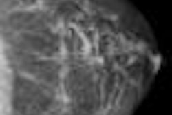
With the SonoCiné system, a transducer from a conventional ultrasound scanner is attached to SonoCiné's computer-guided mechanical arm, which acquires images in longitudinal rows (transverse images), overlapping 7 to 10 mm to ensure complete coverage, according to the researchers. The system's software then creates a cine loop of the images for interpretation. The system was cleared by the U.S. Food and Drug Administration in October 2008.
For the study, SonoCiné was used with multifrequency transducers within at least the 7- to 12-MHz range on several different ultrasound scanners, including the iU22 (Philips Healthcare, Andover, MA), Acuson Sequoia (Siemens Healthcare, Malvern, PA), Logiq 9 (GE Healthcare, Chalfont St. Giles, U.K.), and HDI 5000 (Philips).
One of 10 radiologists with at least 10 years of experience in breast ultrasound interpreted each whole-breast ultrasound study; the mammograms were interpreted by a single radiologist in that institution's usual manner. Although the radiologists were blinded to the results of the corresponding mammograms or whole-breast ultrasound studies, they did in some cases interpret both exams.
To ensure that the original reader had not missed a mammographically detectable cancer, the researchers retrospectively reviewed the mammograms of all participants with cancers detected by whole-breast ultrasound. They also reviewed the mammograms and whole-breast ultrasound studies of all participants with clinically detected interval cancers occurring within one year of a normal mammogram.
Sensitivity and detection rates of automated whole-breast ultrasound
|
There were 57 cancers in 56 study participants. The positive predictive value for biopsy was 39% based on mammography findings, compared with 38.4% for automated whole-breast ultrasound. When whole-breast ultrasound findings were added to mammography, the number of detected invasive cancers 10 mm or less in size tripled from seven to 21, according to the authors. Cancer detections for invasive tumors measuring 11 to 20 mm increased from eight to 14 with the addition of automated whole-breast ultrasound.
In other findings, whole-breast ultrasound detected 32 of 49 cancers (65%) in women with dense or extremely dense breasts, compared with 19 of 49 (39%) by mammography alone (p = 0.02). By adding whole-breast ultrasound to mammography, cancer detections more than doubled, from 19 to 39.
"Limiting [automated whole-breast ultrasound] examinations to a high-risk group with dense breasts, similar to the BRCA1/2 studies with MRI, would dramatically reduce the cost per cancer diagnosis," the authors wrote. "In our study 87% of cancer detections added by [whole-breast ultrasound] were found in the 68% of studies in women with dense/very dense breasts."
Further research should focus on better defining the combination of risk factors and imaging characteristics that warrant supplemental use of automated whole-breast ultrasound, the authors concluded.
By Erik L. Ridley
AuntMinnie.com staff writer
September 10, 2009
Related Reading
Fuzzy 3D ultrasound CAD sharpens breast cancer sensitivity, September 3, 2009
Automated breast US unit not ready to replace handheld, study finds, March 16, 2009
Ultrasound, MRI perform well in dense breasts, March 8, 2009
3D Doppler evaluation helps identify malignant breast lesions, November 6, 2008
Screening US boosts cancer detection rate in high-risk women, May 13, 2008
Copyright © 2009 AuntMinnie.com