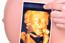
Limiting detailed second-trimester ultrasound exams to patients with risk factors may result in many fetal anomalies going undetected -- and current U.S. reimbursement policy may need updating to reflect this finding, according to research published in the August issue of the Journal of Ultrasound in Medicine.
A research team led by Dr. Stephen Chasen of Weill Cornell Medical College in New York City found that more-detailed ultrasound scans usually reserved for at-risk women found 40% of fetal abnormalities in normal women that would not have been detected on more basic scans (J Ultrasound Med, August 2009, Vol. 28:8, pp. 1015-1018). U.S. reimbursement policy that restricts the more-detailed scans to at-risk women may need to be revised to cover more women.
"It is clear that a policy limiting the structures to be examined in all pregnant women will lead to a substantial proportion of major anomalies going undetected," Chasen said.
The basic scan
Under current clinical practice, women with no risk factors for abnormal pregnancy receive a basic second-trimester ultrasound examination (current procedural terminology [CPT] code 76805), consisting of a survey of intracranial, spinal, and abdominal anatomy; evaluation of the heart; and assessment of the umbilical cord insertion site.
More-detailed scans have been reserved for at-risk women under CPT code 76811, which was introduced in 2003. This study includes the same components of the basic scan, as well as a detailed anatomic evaluation of the fetal brain, ventricles, face, heart, outflow tracts, chest anatomy, and specific abdominal organ anatomy, along with the number, length, and architecture of limbs, according to the authors.
Seeking to evaluate the effects of restricted versus routine use of detailed second-trimester sonography, the Cornell researchers reviewed the records of singleton pregnancies undergoing evaluation at their institution after 14 weeks' gestation from 2004 to 2008. A detailed examination (CPT code 76811) was routinely performed on all patients.
Major structural abnormalities were categorized based on whether the structure would be included in a basic examination (CPT code 76805). In addition, the researchers identified risk factors for anomalies.
Of the 22,335 patients in the study, 218 (1%) had major anomalies. Seventy-five patients (34.4%) elected to have an abortion following the diagnosis.
"There were no significant differences in maternal age, the proportion of pregnancies with risk factors for structural anomalies, or the rate of abortion based on whether the abnormalities involved structures included in the basic examination," the authors wrote.
In 88 patients (40.4%), the abnormal structures would not have been included in a basic examination. These anomalies included 35 clubfoot deformities, 14 cardiac outflow tract anomalies, 10 cleft lip and/or palate cases, seven cases with multiple abnormalities, six arthrogryposis cases, six with limb reduction defect, three with ambiguous genitalia, three with facial teratoma, three skeletal dysplasia cases, and one case of fetal warfarin syndrome.
Sixty-two patients (28.4%) among the 218 with major anomalies were noted to have risk factors for structural anomalies. In 20 of these patients, however, the risk factors were only identified by reviewing medical records and were unknown at the time of the sonographic examinations, according to the study team.
Restricting evaluation of fetal anatomy to those structures included in the basic examination would have prevented detection of a substantial proportion of anomalies, according to the researchers.
"Although these include anomalies that are usually associated with good outcomes, such as clubfoot deformity, anomalies associated with major morbidity, such as cardiac outflow abnormalities, skeletal dysplasia, and arthrogryposis, would also go undetected," the authors wrote.
If a detailed scan had been performed only on the 62 patients with risk factors, and anomalies of structures not included in the basic scan were missed in the remaining patients, 71.6% of anomalies would have been identified with routine use of detailed sonography, according to the authors. Also, if only the 42 patients with known risk factors at the time of the study had received a detailed scan, 66.5% of anomalies would have been identified with routine use of detailed sonography.
By Erik L. Ridley
AuntMinnie.com staff writer
August 4, 2009
Related Reading
Meeting the challenge of obstetrical sonography coding, August 27, 2008
Delivery date change after ultrasound linked with fetal growth restriction, April 14, 2008
Echocardiography parameters offer screening tool for pregnancy complications, March 5, 2008
Study reinforces V/Q, PIOPED II guidelines for PE during pregnancy, February 15, 2008
Fetal anatomic survey yields unexpected anomalies in antepartum US, August 24, 2007
Copyright © 2009 AuntMinnie.com