A sizeable series of patients who underwent virtual colonoscopy after failed colonoscopy revealed significant extracolonic findings that colonoscopy didn't get to.
The data indicate that VC (virtual colonoscopy, or CTC, CT colonography) is a reliable exam for this purpose, and certainly better than double-barium contrast enema, which has limited sensitivity and cannot be performed on the same day as conventional colonoscopy. The retrospective study was conducted by researchers at Beth Israel Deaconess Medical Center in Boston using data acquired at two centers in Israel.
Among radiologists and gastroenterologists alike, VC follow-up after failed colonoscopy is often seen as the most obviously useful indication for virtual colonoscopy. In recent years in the U.S., an increasing number of local Medicare payors, as well as a handful of private insurers, have begun paying for this indication. Only a few studies have tracked the use of VC after failed colonoscopy, however, since a report by Dr. Martina Morrin and her colleagues from Beth Israel in 1999 (American Journal of Roentgenology, April 1999, Vol. 172:4, pp. 913-918).
The present study, though performed on four-slice CT scanners with data acquired between 1999 and 2002, showed that VC can reliably increase the diagnostic yield in portions of the colon not visualized during incomplete colonoscopy (Radiology, August 2007, Vol. 244:2, pp. 471-478).
The U.S.-approved alternative -- double-contrast barium enema -- is less advantageous, wrote study authors Dr. Laurian Copel, Dr. Jacob Sosna, Dr. Jonathan Kruskal, Dr. Richard Farrell, Dr. Martina Morrin, and their colleagues from the Sackler School of Medicine in Zerifin, Israel, Hadassah Hebrew University Medical Center in Jerusalem, and Beth Israel Deaconess Medical Center in Boston.
"When colonoscopy is incomplete, double-contrast barium enema examination has been performed to complete the inspection of the nonvisualized part of the colon," they wrote. "However, double-contrast barium enema examination can be associated with considerable patient discomfort, and may not be tolerated on the same day as colonoscopy. Furthermore, data from the National Polyp Study Work Group, in which comparison was made between paired colonoscopy and double-contrast barium enema examination, indicated that the latter depicted only 48% of adenomas greater than 10 mm in diameter, 53% of adenomas 6-9 mm in diameter, and 32% of adenomas 5 mm or smaller in diameter."
The researchers sought to retrospectively assess the positive predictive value of VC in patients referred for further examination after incomplete colonoscopy.
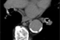
VC after incomplete colonoscopy
During the three-year study period, 546 VC exams were performed at the Israeli centers due to incomplete colonoscopy by gastroenterologists who had at least five years experience performing colonoscopy.
The main reason for incomplete colonoscopy was redundant or tortuous loops, which occurred in nearly 40% of the cases. The most frequent location of incomplete colonoscopy was the sigmoid colon, in 23.8%. Of the 546 patients, 492 (90.1%) were considered at high risk for developing colorectal cancer owing to symptoms or a personal or family history of polyps or cancer, whereas 54 patients (9.9%) were considered a low-risk screening population.
The patients underwent cathartic bowel cleansing, most (456/546) with two 45-mL doses of sodium phosphate (Fleet Prep Kit 1; Fleet Pharmaceuticals, Lynchburg, VA) along with 10 mg of bisacodyl. Following room air insufflation to patient tolerance, and the acquisition of scout images to check insufflation, the patients were scanned on either a four-slice (n = 512) or eight-slice (n = 34) scanner (LightSpeed QX/i or LightSpeed Ultra, respectively, GE Healthcare, Chalfont St. Giles, U.K.).
The four-section scans were acquired with a slice thickness of 2.5 mm (n = 162) or 1.2 mm (n = 350), while the eight-section scans were acquired at 1.25-mm slices (n = 34), with all scans at 100-200 mAs and 120 kVp. The data were interpreted on a GE Advantage Windows workstation by 14 radiology fellows, then by three abdominal radiologists experienced in VC interpretation, who were blinded to the initial results.
For the study, two VC-experienced radiologists reviewed the VC and colonoscopy results in order to identify all endoscopically nonvisualized lesions 6 mm or larger, segment by segment.
"By using subsequent colonoscopic and postcolonoscopic surgical findings (in 45 patients) as the reference standard, we determined whether lesions previously seen at CT colonography represented true-positive or false-positive findings," Copel et al explained. A true-positive match had to be in the same or an adjacent segment, and the size correlated within 50% of the original diameter. All studies were evaluated on a GE Advantage workstation.
The results showed that in 72 (13.2%) of patients, VC depicted 88 endoscopically nonvisualized lesions 6 mm or larger. Eleven patients were reported to have 12 masses 20 mm or larger at subsequent colonoscopy, but one patient had no mass.
"Eighteen patients had 23 large (10-19-mm) polyps they were suspected of having, and 47 patients had 53 medium (6-9-mm polyps) that they were suspected of having," the authors reported.
Seventy-two patients underwent follow-up colonoscopy at a median of 31 months (range 6-42 months) due to their CTC findings. Optical colonoscopy was repeated in 100% of cases for masses, 94% for large polyps, and 45% for medium-size polyps, they wrote.
The per-patient positive predictive values for virtual colonoscopy for masses, large polyps, and medium polyps were 90.9%, 91.7%, 64.7%. Per lesion positive predictive values were 70%, 33.3%, and 30.4%, respectively.
Incomplete colonoscopy has been reported at rates ranging from 2% to 25% in various studies, and the rate in the present series was 3.5%, the authors reported.
"Although the capability of CT colonography to depict polyps is both operator and technique dependent, this modality has a relatively high specificity," the authors wrote. "Our study findings indicate that our clinicians appear to be comfortable in accepting the negative results of a CT colonographic examination -- to the best of our knowledge, none of the 474 patients (86.8% of the cohort) in our study whose CT colonographic findings were reported as normal underwent repeat colonoscopy for examination of the nonvisualized colon during the follow-up period. By contrast, 96% of patients underwent repeat colonoscopy if CT colonography depicted endoscopically nonvisualized lesions of 10 mm or larger in diameter."
VC finds lesions not reachable by colonoscopy
VC correctly depicted 11 masses larger than 20 mm that were not seen endoscopically, and in six patients, seven colonic carcinomas in the endoscopically nonvisualized part of the colon were correctly identified at VC. The positive predictive value of 91.7% for masses was similar to other reports. Only one 30-mm cecal lesion found at VC was not confirmed at repeat colonoscopy -- it appears that a large but normal fold in the cecum was mistaken for a mass, the group wrote.
Slightly more than half of the cases with medium-sized polyps (6-9 mm) were followed up, owing to a tendency by the gastroenterologist to concentrate their efforts on larger, higher-risk lesions. The low follow-up rate for these lesions was cited as a study limitation, however. The study was retrospective as well, and there was selection bias inasmuch as 90% of the patients were at elevated risk of colorectal polyps and cancer.
In all, 19 patients had 23 polypoid lesions (including seven 10 mm and larger) reported at VC that were subsequently proven to be false positive at repeat colonoscopy, the authors noted. Yet a second review of the images by experienced radiologists found that most of them [17 of 23 (74%)] would have been reported again as polypoid lesions.
Lacking the benefit of segmental unblinding of colonoscopy results, "one must allow for the possibility that the endoscopist that performed the repeat colonoscopy was not always able to accurately identify the location of the polypoid lesion," the authors wrote. "We suggest that, when a year has passed and no colonoscopy has been performed for medium polyps that are suspected, it might be reasonable to send a reminder to the referring physician about the findings, especially in patients suspected of having 8-9 mm polyps."
Some 96% of patients with VC-detected lesions 10 mm and larger underwent repeat colonoscopy, the authors noted.
"Our study findings indicate that CT colonography has the potential to become an accepted technique for the evaluation of the nonvisualized part of the colon after incomplete colonoscopy, and that it can increase the diagnostic yield of masses and clinically important polyps in this part of the colon," they wrote.
By Eric BarnesAuntMinnie.com staff writer
August 8, 2007
Related Reading
Sex, age, location influence incomplete colonoscopy rates, July 3, 2007
Colonoscopy more often successful in mornings, June 22, 2007
Study: VC is cost-effective in efficient exam settings, May 9, 2007
Screening model calls VC most cost-effective colon exam, April 24, 2007
Higher costs seen in post-VC extracolonic follow-up, November 3, 2006
Copyright © 2007 AuntMinnie.com