Sonography remains an effective tool for evaluating the acute abdomen in pediatric patients, according to a review article in the Journal of Ultrasound in Medicine. Thanks to its noninvasive nature and sensitivity in finding lesions that produce acute abdominal disease, ultrasound is most often the imaging procedure deployed after an initial supine and horizontal-beam radiograph.
Causes of the acute abdomen vary by age. In the premature neonate, sonography may be useful when perforation and abscess formation is suspected, wrote Dr. Diane Babcock of Cincinnati Children's Hospital Medical Center (JUM, August 2002, Vol.21:8, pp. 887-899).
For full-term neonates, sonography can aid in the diagnosis of small-bowel obstruction, particularly in the gasless abdomen by confirming the presence and location of distended, fluid-filled bowel loops. In addition, reversal of the superior mesenteric artery and vein positions can suggest malrotation.
Intussusception is a particularly common cause of acute abdomen in infants. This can be seen on ultrasound as a mass with a typical "target" appearance on a cross section, due to multiple layers of bowel and a pseudokidney appearance on longitudinal scans, Babcock wrote. Negative sonographic findings can exclude intussusception with nearly 100% accuracy.
Doppler sonography also does a good job of evaluating bowel ischemia and perforation risk in infants. Sonography of the scrotum and inguinal area can also confirm an inguinal hernia, which may incarcerate and appear as an acute abdomen, Babcock stated.
For children with upper right quadrant pain, sonography can demonstrate gallbladder wall thickening and tenderness when scanning is performed over the gallbladder. Renal diseases, such as acute dilatation, can cause an acute abdomen, and ultrasound can demonstrate dilatation of the collection system, according to the article.
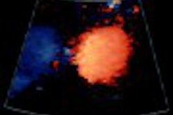
Left upper quadrant pain can be caused by spleen disorders, such as splenic torsion, which is characterized on ultrasound as an abnormal spleen location in the mid or lower abdomen and decreased flow on Doppler imaging, Babcock wrote. Splenic infraction may produce abnormal echogenicity or areas of abnormal perfusion on Doppler imaging.
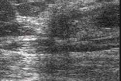
Right lower quadrant pain can often mean acute appendicitis, the most common acute surgically treated ailment in infants and children. Although imaging is used only in atypical or uncertain appendicitis cases, the appendix can be viewed via ultrasound with graded compression of the right lower abdomen, the article noted.
Doppler imaging may aid in the diagnosis, as an acutely inflamed appendix has increased flow compared with normal bowel, she noted. And ultrasound can also be helpful in imaging other causes of right lower quadrant pain.
"Inflammatory bowel disease and gynecological disorders may also cause pain in the right lower quadrant, and sonography is particularly useful in girls to distinguish appendiceal disease from gynecologic disorders," Babcock wrote.
For midabdominal pain, ultrasound can reveal thick-walled bowel loops and small-bowel intussusception from bleeding disorders that produce hemorrhage into the bowel wall. Patients without bleeding disorders but with unusual-appearing masses and hematoma could be victims of child abuse, she said. Sonography can also reveal enlarged mesenteric lymph nodes and can exclude other conditions, such as an inflamed appendix or acute dilatation of the renal pelvis.
For imaging of gynecologic disorders, ultrasound is useful in demonstrating the normal or abnormal ovary and uterus, abnormal fluid collections, and masses, Babcock said. For example, a hemorrhage into a follicular cyst is revealed as an ovarian mass without internal flow.
In nonsurgically treated cases of abdominal pain in children, sonography can rule out other organic causes of abdominal pain. Sonography may also occasionally diagnose pneumonia involving the lower lobes, by serendipitous visualization of an abnormal nonaerated lung above the diaphragm.
"Sonography in conjunction with color and pulsed-Doppler imaging is a valuable tool in the evaluation of the acute abdomen in the pediatric patient," Babcock concluded.
By Erik L. RidleyAuntMinnie.com staff writer
October 4, 2002
Related Reading
Emergency Imaging of the Acutely Ill or Injured Child, September 10, 2002
SPECT coregistration improves treatment at pediatric care center, June 18, 2002
Ultrasound disappoints in abdominal trauma, December 14, 2001
Copyright © 2002 AuntMinnie.com