(Ultrasound Review) Dynamic sonography in the hands of experts provides valuable information about disk displacement within the temporomandibular joint (TMJ), according to researchers at the University of Innsbruck in Austria.
In a study published in the American Journal of Roentgenology, they established that ultrasound imaging was able to demonstrate internal derangement, and disk displacement with and without reduction. Internal derangement is defined as an abnormal position of the articular disk relative to the mandibular condyle and the articular eminence.
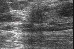
Sonography was performed on the right and left TMJ in 64 consecutive patients in the closed-mouth and maximal open-mouth positions, and during opening and closing.
"The orientation of the scanning was based on a standardized protocol to obtain cross-sections intersecting the anterosuperior joint compartment in a sagittal-to-frontal plane," they reported. With the patient supine, the transducer was placed along the long axis of the mandibular ramus over the TMJ and zygomatic arch.
According to the authors "the bony landmarks of the mandibular condyle and the articular eminence are visualized as hyperdense lines. We identified the course of the disk’s motion by having the patients slightly move the mandible." They emphasized that the transducer orientation must be maintained during jaw movement to avoid artifactual changes in disk appearance, in particular echogenicity.
"In evaluating findings of the closed mouth, we considered the position of the disk to be normal if the intermediate zone of the disk was located between the anterosuperior aspect of the condyle and the posteroinferior aspect of the articular eminence. Disks with the intermediate zone located anterior to this position were considered displaced," they indicated.
There were 87 internal derangements in all. Using MRI as the gold standard, dynamic sonography correctly diagnosed internal derangement in 93% of cases, disk displacement with reduction in 82% of cases, and displacement without reduction in 83% of cases. They established that "the accuracy of prospective interpretation of high-resolution sonograms of internal disk displacement with reduction, and disk displacement without reduction was 95%, 92%, and 90%, respectively."
Disk displacement of the temporomandibular joint: sonography versus MR imagingR Emshoff et al
Department of oral and maxillo-facial surgery, University of Innsbruck, Innsbruck, Austria
AJR 2002 June; 178:1557-1562
By Ultrasound Review
July 22, 2002
Copyright © 2002 AuntMinnie.com