(Ultrasound Review) According to radiologists at the University of Michigan, color Doppler ultrasound provides information that enables discrimination between normal prostatic tissue and cancer.
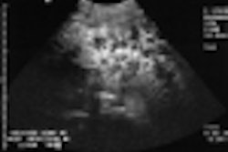
They analyzed 3-D images and demonstrated that color pixel density was the most promising measure. An article outlining their findings was published in the July issue of the Journal of Ultrasound in Medicine.
"The rationale for using color flow imaging is that cancer will develop an increased blood supply by angiogenesis, and this can show up as increased and abnormal color flow patterns in and around the cancerous regions," the authors stated.
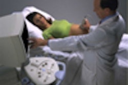
Thirty-nine patients had transrectal ultrasound examination prior to radical prostatectomy. Using a 9–5 MHz tightly curved array transducer oriented in a sagittal plane, the probe was rotated from right to left through the entire gland at increments of 0.540.
Each color and grayscale image was digitally stored, and the appearances were compared with corresponding histologic sections. A special apparatus was utilized that enabled acquisition of images "in a stable, reproducible way so that scan slices can later be reconstructed in the correct geometric configuration," the authors stated.
Comparisons were made between the presence and location of prostate cancer and microvessel count. Color pixel density provided an accuracy of 80% in diagnosing tumor tissue and, when combined with normalized mean Doppler power, the accuracy increased to 84%. The latter measurement took into account the variations in normal prostate vascularity between patients.
"The two simple components of Doppler ultrasonography that contribute to discriminating prostate cancer from undiseased prostate are color pixel density and normalized mean power in color pixels," the authors concluded.
"Analysis of 3-D Doppler ultrasonographic quantitative measures for the discrimination of prostate cancer"Aaron P Moskalik et al
Dept of Basic Radiological Sciences, University of Michigan, 200 Zina Pitcher Pl, Kresge III, Room 3316, Ann Arbor, MI 48109-0553, USA
J Ultrasound Med 2001(July); 20:713–722
By Ultrasound Review
August 9, 2001
Click here to post your comments about this story. Please include the headline of the article in your message.
Copyright © 2001 AuntMinnie.com