A pair of new studies suggests that cardiac imaging still has plenty to gain from echocardiography -- specifically, from its strong predictive value in infective endocarditis to its accurate assessment of patients with suspected acute coronary syndrome (ACS).
Real-time myocardial contrast echocardiography (RTMCE) has enabled fast and accurate bedside evaluation of patients with suspected ACS, according to cardiologist Dr. Thomas R. Porter and colleagues from the University of Nebraska Medical Center in Omaha.
"Our results demonstrated that dobutamine infusion with RTMCE was both safe and effective" in gauging which emergency patients are at risk for recurrent events, the researchers wrote in the latest International Society of Cardiovascular Ultrasound publication (Echocardiography, July 2005, Vol. 22:6, pp. 487-494).
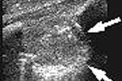
Meanwhile, in the latest American Heart Association journal, cardiologist Dr. Gilbert Habib from the Hôpital de la Timone in Marseille, France, and co-investigators from other French and Italian institutions report on the largest prospective evaluation of vegetations or clots in infective endocarditis (IE).
Their findings confirmed that large vegetations of greater than 10 mm and severe vegetation mobility as seen on echocardiography are both associated with an increased risk of embolic events (Circulation, July 5, 2005; Vol. 112:1, pp. 69-75).
Despite recent improvement in diagnostic and therapeutic strategies, the authors noted, IE is associated with in-hospital mortality ranging from 16% to 25%. Embolic events (EE) are reported in 13% to 49% of cases.
"However, previous studies that attempted to identify baseline predictors of mortality and embolism led to conflicting results," the authors wrote. Therefore, they designed a trial of systematically using transesophageal echocardiography (TEE) to assess the predictive value of various parameters.
Consecutive patients referred to four European centers over a 10-year period ending in 2003 with a definite diagnosis of IE based on the Duke University criteria were included in the study. Those with pacemaker IE were excluded, generating a study group of 384 patients.
Transthoracic echocardiography (TTE) and TEE were performed in all cases. Vegetations could be seen in 320 patients (83%) using TEE but in only 192 patients (50%) by TTE.
Measurements of vegetation length were performed in various planes, with the maximal length of the largest of multiple vegetations used for analysis. Clot mobility was characterized as being absent, low, moderate, or severe -- the last described as "prolapsing vegetation that crosses the coaptation plane of the leaflets during the cardiac cycle."
"Along with baseline clinical and microbiological features, the assessment of vegetation characteristics (length and mobility) allows identification of patients who are at highest risk for new EE and death," the authors wrote.
Specifically, a vegetation longer than 15 mm was a predictor of one-year mortality with an adjusted relative risk of 1.8 (p = 0.02), they reported. Overall, more than 20% of the patients died within one year of diagnosis.
Vegetation length greater than 10 mm predicted an embolic event with an adjusted odds ratio of 9 (p = 0.004), and severe vegetation mobility did likewise with an adjusted odds ratio of 2.4 (p = 0.04).
"[T]he fact that very large vegetations were independently associated with a worse prognosis is not surprising because this feature is frequently associated with severe valve destruction and a high embolic risk," the authors noted.
However, "among the patients with vegetation length >15 mm, 34% had no other indication for surgery and would have been identified as high-risk patients by echocardiography before standard indications for surgery were met."
"Thus, the measurement of vegetation length at the time of diagnosis of IE is strongly recommended as part of the initial risk stratification," they concluded. "Patients with the largest vegetations should be considered at high risk for subsequent serious complications."
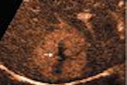
In the second study, the Nebraska investigators looked at the new technique of dobutamine stress RTMCE, which allows for simultaneous perfusion and wall motion analysis.
Their goal was to compare the myocardial perfusion analysis (MPA) with the contrast-enhanced wall motion analysis (WMA) for detecting coronary artery disease (CAD) in emergency department patients, and to gauge the prognostic value of both parameters in patients with possible ACS.
The researchers enrolled 158 consecutive patients who presented to their emergency department with chest pain between January 2000 and May 2003. Excluded were those with persistent electrocardiographic ST segment elevation in at least two leads, elevated serial values of creatine kinase myocardial band (CKMB) characterizing acute myocardial infarction, severe uncontrolled hypertension, severe valvular disease, pregnancy, major ventricular arrhythmias, or contraindications to any drug used in the study.
RTMCE was performed on an HDI 5000 and Sonos 5500 scanner (Philips Medical Systems, Andover, MA) and a Sequoia 6.0 scanner (Siemens Medical Solutions, Malvern, PA). Two contrast agents were used in the study: Optison (Mallinckrodt, St. Louis, MO) in 133 patients and Definity (Bristol-Myers Squib, North Billerica, MA) in 25.
"MPA was considered positive for ischemia when ≥ 2 contiguous segments failed to exhibit contrast enhancement at peak stress as compared with other segments at the same depth in the same view, as well as compared to contrast enhancement in the same segments at baseline using a side-by-side image analysis," the authors explained.
"Since these patients were examined within the first 48 hours of presenting with a potential ACS, WMA was considered positive for ischemia when ≥ 2 contiguous segments exhibited resting or inducible wall motion abnormalities," they continued.
Sixty-one (39%) patients underwent quantitative coronary angiography within one month of their RTMCE, enabling the researchers to gauge the echo technique's success in predicting the significant CAD found at angiography.
"MPA by RTMCE improved the sensitivity, negative predictive value, and accuracy for detection of CAD when compared with WMA, while maintaining similar specificity," the authors wrote, summarizing their results.
In particular, MPA achieved 92% sensitivity compared to WMA's 62%, and 77% specificity versus 85%. Both achieved a 94% positive predictive value while MPA's negative predictive value was much higher at 71% versus 38%. Overall accuracy for MPA was 88% versus 67% for WMA.
The researchers also evaluated the 131 patients who could be followed up for a median of 16 months, and found that 25 (19%) sustained subsequent cardiac events.
"Age-adjusted multivariate analysis demonstrated that a positive MPA (hazard ratio = 3.23; 95% CI = 1.23-8.49; p = 0.018) and male sex (hazard ratio = 3.29; 95% CI = 1.21-8.97; p = 0.02) were the only independent predictors of all cardiac events," they wrote.
Their results demonstrated that dobutamine infusion with RTMCE was both safe and effective in evaluating these patients within 48 hours after presenting to the emergency department, the researchers stated.
"MPA and WMA were highly predictive of outcome in these patients and had excellent negative predictive value," the authors concluded. "However, MPA was able to add to the predictive value of wall motion by identifying patients with normal wall motion responses to dobutamine who were still at risk for cardiac events."
By Tracie L. Thompson
AuntMinnie.com staff writer
July 15, 2005
Related Reading
Multislice CT reveals previous plaque rupture in coronaries, June 14, 2005
S. aureus endocarditis has worse outcomes than other bacterial endocarditis, June 29, 2005
Cardiac MRI useful for predicting significant coronary artery disease, December 20, 2004
Real-time MR stress imaging detects cardiac wall-motion abnormalities, September 23, 2002
Echocardiography overused in work-up of infective endocarditis, August 13, 2002
Multiple plaque ruptures identified during acute coronary syndromes, July 23, 2002
Copyright © 2005 AuntMinnie.com