Hormonal variations in breast vascularity should be considered when using ultrasound to evaluate breast lesions, according to research published in the January issue of the Journal of Ultrasound in Medicine.
To determine if hormonal changes during the course of a menstrual cycle yield sonographically detectable variations in breast glandularity and vascularity, a team of researchers from the University of Pennsylvania Medical Center in Philadelphia and Crozer-Chester Medical Center in Upland, PA, performed ultrasound studies on seven premenopausal women (JUM, January 2005, Vol. 24:1, pp. 67-72).
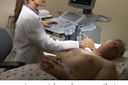
Breast ultrasound was performed on the subjects every three days during the work week for one menstrual cycle, imaging in multiple planes using color Doppler, power Doppler, and B-scan modes on a Sonoline Elegra system (Siemens Medical Solutions, Malvern, PA) with a 10- to 13-MHz transducer.
The studies were performed by the same radiologist and at the same time of the day for each patient, with the same regions being scanned each time. The seven patients produced 49 scans, and 30 images were digitized for each scan. A total of 1,500 images were analyzed for the entire study; the images were recorded on tape and analyzed for the area of perfusion, blood volume, and flow rate.
The researchers then digitized the grayscale (B-scan), color, and power Doppler images, which were analyzed for image brightness and color-level change by software developed at the University of Pennsylvania Medical Center.
Vascularity was measured by mean color level, area covered by color pixels, and color-weighted fractional area. Progesterone and estradiol levels were measured from saliva collected from the patients.
Two of the seven volunteers were taking oral contraceptives (OCPs) at the time of the study. Of the five patients who were not taking OCPs, four had peaks in vascularity correlating with midcycle peak hormone levels, according to the researchers.
Vascularity increases ranged from 50% to 320% from the baseline level on power Doppler sonography. With color Doppler, three of five patients also had peaks in vascularity over the course of the cycle, the authors reported. Increases from baseline levels ranged from 60% to 190%.
In the two patients taking OCPs during the study, no significant peaks in vascularity were seen over the course of the menstrual cycle.
"Our preliminary experience showed that variations in the vascularity of the breast tissue occurred over the course of the menstrual cycle in patients who were not taking OCPs," the authors wrote. "This variation may be dramatic, with a change of as much as 320% above baseline levels."
By Erik L. Ridley
AuntMinnie.com staff writer
January 13, 2005
Related Reading
Tumor type, breast profile determine value of mammo, US, and MR, January 6, 2005
Breast US readers need more practice with postop changes, December 22, 2004
Breast imagers fare better with hands-on approach to screening ultrasound, December 17, 2004
ACRIN updates multiple breast cancer trials, April 15, 2004
Copyright © 2005 AuntMinnie.com
