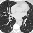
By modulating x-ray tube output based on patient characteristics, instead of using a fixed tube current, Texas researchers were able to lower radiation dose in pediatric CT studies by 23%, according to a poster presentation at this week's American Association of Physicists in Medicine (AAPM) meeting.
Thanks to improved treatments, the outlook for children diagnosed with cancer is more positive than ever, and the majority of children survive their diseases. But their future is not entirely bright, because childhood cancer survivors have higher than average risks of developing a second cancer later in life.
The reason for this is still unknown, but oncologists attribute this development of additional cancers to genetic predisposition, radiation therapy treatments, chemotherapy -- and also to radiation dose exposure from diagnostic imaging exams. Repeated exposure to high radiation doses from CT exams used to monitor the effectiveness of treatment is a much-discussed subject by pediatric oncologists.
This problem is being addressed by medical physicists at M. D. Anderson Cancer Center at the University of Texas in Houston, who have been working to reduce the risk of repetitive CT scan exposure. In their AAPM poster, medical physicists described how they were able to reduce radiation exposure by 23% without compromising the quality of patient care.
Dianna Cody, PhD, a professor in the department of imaging physics, said that she and colleagues used a logical three-step procedure implemented between October 2008 and November 2009 to reduce radiation as measured by dose-length product (DLP) by 28.6%, on average.
At M. D. Anderson, all 13 CT scanners in use can modulate radiation output based on the amount of tissue being scanned, ramping up in areas of relatively thick tissue and tamping down in thinner areas such as the neck. Cody and colleagues programmed the CT scanners to produce a specified image quality while automatically adjusting the radiation dose up or down.
The physicists established tube-length current modulation protocols in October 2008 based on the recommendations of Donald Frush, MD, chief of pediatric radiology at Duke University Medical Center in Durham, NC. The largest radiation dose reduction was achieved after replacing a fixed tube current technique with this one.
In August 2009, they subsequently increased the noise index by a factor of two, and in November, they lowered the minimum and maximum tube current (mA) values by 20%. Pediatric radiologists were consulted at each stage to verify that image quality was adequate and patient care was not compromised, Cody explained.
Four chest CT scans taken of 15 patients over time were analyzed to evaluate dose reduction. The physicists developed a computer program to calculate effective mAs, CT dose index volume (CTDIvol), and dose-length product for each of the four exams per patient. The program calculated effective mAs by averaging all the mA values used during the scan, multiplying the average mA by the rotation time(s), and then dividing the current-exposure time product (mAs) by the pitch.
The weighted CTDI in units of mGy/mAs multiplied by the effective mAs was used to calculate CT dose index volume. The dose-length product was calculated by multiplying the CT dose index volume by the scan extent, defined as the difference between the patient start and stop positions as they appeared in patient images.
The average effective mAs dropped from 146 mAs at baseline to 91 mAs after implementing all three strategies, a total of 33%, on average, Cody reported. The average CTDIvol dropped from 38.0 mGy to 25.3 mGy, and the average DLP dropped from 848 mGy-cm to 544 mGy-cm. This represented a total average decrease of 31.1% and 26.6%, respectively. However, not all of the 15 patients experienced the dose savings expected, because interval growth in some instances warranted an increase in technique, according to Cody.
She and her colleagues are currently evaluating adaptive statistical iterative reconstruction (ASIR), organ shielding, and other approaches as means of achieving even greater dose savings for pediatric cancer patients undergoing CT exams, Cody told AAPM attendees.
By Cynthia E. Keen
AuntMinnie.com staff writer
July 21, 2010
Related Reading
Cancer survivors have higher death risk for decades, July 14, 2010
Child cancer survivors have higher heart risk, December 10, 2009
Risk of breast cancer after radiotherapy in childhood quantified, July 21, 2009
Copyright © 2010 AuntMinnie.com


