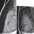
ARLINGTON, VA - MRI is superior to mammography for evaluating the response to neoadjuvant chemotherapy for breast cancer, according to the early results of a trial from the University of California, San Francisco (UCSF) and nine other academic centers in the U.S. The results were presented at last week's American College of Radiology Imaging Network (ACRIN) fall meeting.
Precisely how much MRI improves neoadjuvent chemotherapy response will have to wait until final data are released later this year. And although early results are promising, data on whether MRI after the first cycle of chemotherapy can predict disease-free survival are not expected until mid-2009.
The study was developed under ACRIN's protocol 6657, representing the imaging side of the larger I-SPY (Investigation of Serial Studies to Predict Your Therapeutic Response With Imaging and Molecular Analysis) trial aimed at gauging breast cancer treatment response. Sponsored by the Cancer and Leukemia Group B Foundation (CALGB), the I-SPY trial is "testing imaging and tissue-based biomarkers in combination, predicting neoadjuvent response to standard chemotherapy," explained Nola Hylton, Ph.D., a principal investigator from UCSF who discussed the results last week.
For the combined studies, a network of oncologists examined 216 eligible women (median age, 49; range, 27-68) with locally advanced breast cancers 3 mm or larger in diameter who would undergo neoadjuvant anthracycline-based chemotherapy, followed optionally by taxane.
"MRIs and core biopsies were acquired at multiple points during treatment" and addressed whether "MRI measurements of treatment response predict three years of disease-free survival, and secondarily, can MR measurements after one cycle of treatment provide an early prediction of response," Hylton said.
Participants were scanned four times during chemotherapy, including once pretherapeutically, once after the first cycle of chemotherapy, and a third time between the anthracycline and Taxol agents. A fourth scan prior to surgery was intended to detect residual disease and evaluate the post-treatment sensitivity and specificity of MRI. Mammography scans were acquired to coincide with the first and last MRI scans, she said.
MRI measurements included morpholgic measurements of the tumors classified according to BI-RADS criteria for breast MRI, including tumor diameter measurements, Hylton said. "We're also measuring by computer the volume of the tumor and the microvascular parameters of the tumor, PE [percent enhancement] and SER [signal enhancement ratio]," used to distinguish malignant tissue, she said.
The investigational software assesses tumor volume quantitatively based on functional rather than anatomic criteria, she explained. "We are measuring something that is based on how these tumors enhance, and assigning [volume] based on an algorithm calling it part of the tumor or not. So it's really a virtual volume [that defines] areas of the image based on function, in this case how the tumors enhance."
The investigators acquired one T1-weighted precontrast and two T2-weighted postcontrast scans, "and from that we looked at the early ratio of enhancement from the early time point to the late time point," she said. This calculation yields the signal enhancement ratio, which distinguishes tumor from nontumor. Another protocol ensures that the direction of diameter measurements remains constant over the course of multiple imaging exams.
At the end of surgery, 43% of the patients were complete responders, 38% were partial responders, and 10% demonstrated stable disease. "There were a larger portion of complete responders among those who also received Taxol," she said. "And there were a total of 82 pathologic complete responders, meaning that there was no invasive disease left at pathology. Sixty percent of patients had a complete solid lesion, and 32% had two identifiable lesions in the breast."
Compliance with the study was surprisingly good, especially considering the complexity of the protocol, requiring both biopsy and multiple imaging exams in addition to treatment, she said. In addition, lesion morphology was very similar to a pilot study. There were 36 single masses; 65 multilobulated masses with well-defined margins; 66 lesions with area enhancement, irregular margins, and nodularity; 30 of the same without nodularity; and 19 patients with septal spread.
"Overall we are seeing what we expected to see, which is better correlation of MRI tumor volume by volumetric measurement of tumor with pathologic size of the disease when compared with other end-of-treatment outcomes such as mammography, clinical exam, or even the measurement of the longest diameter on MRI," Hylton said. "We're seeing better correlation of volume with the clinical response than mammography, clinical exam, or MRI longest diameter, and we are also seeing suggestions that the early volume change is predictive of the overall response to treatment presurgically."
Once the analysis has been completed, more precise data will be presented at the 2008 RSNA meeting in Chicago, she said. The study also is getting an amended protocol and 140 new patients, thanks to research at the University of Minnesota in Minneapolis suggesting that single-voxel MR spectroscopy (MRS) can be used to measure choline levels after the first round of chemotherapy and, potentially, treatment response. The new patients will follow a similar protocol, except that MRS will now be performed along with MRI throughout the course of treatment, Hylton said.
Also in the works is the phase II I-SPY 2 trial, which will test a similar protocol with new chemotherapy agents and new biomarkers. The study is designed so that additional promising chemotherapy agents can be inserted into the protocol to replace less successful therapies. On the imaging side, the MRI volume calculation will be performed in real-time "so that it can be used in the assignment of patients to randomization," Hylton said.
A commercial version of the investigational volumetric software (Radiology, July 2008, Vol. 248:1, pp. 79-87), currently under development, will be installed at each site for real-time volumetry.
The SPY-2 trial "is a wonderful opportunity to look at new functional imaging methods to test them in the setting of novel agents, and to test imaging biomarkers in vivo," Hylton said.
By Eric Barnes
AuntMinnie.com staff writer
October 7, 2008
Related Reading
MRI techniques show early tumor response to radiation, September 30, 2008
Breast MRI can guide radiation therapy to lymph nodes, September 22, 2008
Routine MRI at breast cancer diagnosis linked to treatment delays, September 9, 2008
Breast MRI gains momentum for screening high-risk women, September 2, 2008
Breast MRI develops role as surgical planning tool, September 2, 2008
Copyright © 2008 AuntMinnie.com

