MRI has joined the parade of extracolonic findings detected at virtual colonoscopy, in a study of 375 symptomatic individuals in Germany. Researchers from University Hospital Essen found clinically significant findings in 12% of their cohort.
In addition to the invasiveness and procedural discomfort associated with conventional colonoscopy, "the view of the endoscopist is limited to (assessing) the colonic lumen, and thus the evaluation of extracolonic abdominal organs is not possible," though the presence or absence of extracolonic findings "might be important for further patient management," wrote Dr. Waleed Ajaj and colleagues in European Radiology (January 24, 2007).
The group sought to evaluate the utility of dark-lumen MR colonography (MRC) for assessing extracolonic organs, wrote Ajaj and his colleagues Dr. Mathias Goyen, Dr. Susanne Ladd, and Dr. Guido Gerken from Essen, and Dr. Stefan Rheum from the University of California, Los Angeles.
The patients (249 men, 131 women; ages 18-76, mean age 51.7 years) were all considered to have an above-average risk of colonic disease. They had been referred for MRC for various indications, such as abdominal pain, known Crohn's disease or ulcerative colitis, or a positive fecal occult blood test.
Prior to MRC, all patients underwent a standardized bowel cleansing procedure consisting of 3,000 mL of a polyethylene glycol solution (GoLytely, Braintree Laboratories, Braintree, MA). Scopolamine (40 mg of Buscopan, Boehringer Ingelheim, Ingelheim, Germany) was administered intravenously prior to bowel distension with warm tap water (2,000-2,500 mL) to patient tolerance.
All exams were performed on a 1.5-tesla Magnetom Sonata (Siemens Medical Solutions, Malvern, PA) equipped with a high-performance gradient system. Two surface coils were used in conjunction with the built-in spine array coil, which allowed coverage of the entire colon, according to the authors.
Following the bowel distension enema, a precontrast T1-weighted 3D gradient-echo dataset with integrated fat suppression (VIBE sequence) was acquired in the coronal plane in a single breath-hold of approximately 22 seconds.
The parameters included TR/TE of 3.1/1.1 msec, flip angle of 12°, field-of-view of 450 x 450 mm, matrix of 168 x 265 with an effective slice thickness of 1.5-2.0 mm depending on the patient's size.
Next a paramagnetic contrast agent (gadobenate dimeglumine, MultiHance, Bracco, Milan) was administered intravenously at 0.2 mmol/kg of body weight at 3.5 mL/sec. After a delay of 75 and 120 seconds, respectively, a second and third 3D dataset were acquired using the same parameters.
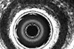
"The lack of contrast enhancement between the pre- and postcontrast scans rules out the presence of a colorectal mass," explained the authors. "The intravenous application of paramagnetic contrast technique allows the direct depiction of the colorectal wall. Thus, the bright colonic wall can be easily discriminated from the dark, water-filled colonic lumen."
Finally a 2D FLASH sequence of the entire abdomen and pelvis was acquired axially using the following parameters: TR/TE of 1.8 msec, flip angle of 70°, and slice thickness of 5 mm.
The data were evaluated on a Virtuoso (Siemens) workstation by two radiologists experienced in MRC by consensus and in a blinded fashion. The results were divided into known and unknown, clinically relevant, and irrelevant findings. When extracolonic findings were suspected, the patients were recommended for further exams.
The exam was successful and well tolerated in the 375 of the 380 patients who decided to go through with the exam, though "in two patients a big part of the water enema was spilled on the scanner table," Ajaj and colleagues wrote. The mean exam time was 31 minutes (20-37 minutes), and image quality was good on a scale of 1 to 3, 1 being good (average 1.4, range 1.0 to 1.8).
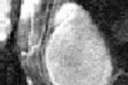
No polyps, carcinoma, or inflammatory signs of the colonic wall were seen in 318 patients (85%). Among the remaining 15%, 47 colorectal lesions (6-25 mm) were found in 39 subjects, and five of these were thought to be malignant. Polyps in three patients were previously known, and signs of Crohn's disease and ulcerative colitis were seen in another 13.
Colonoscopy confirmed the colorectal lesions in the 39 subjects, but histopathology of the five suspected carcinomas found only three of the five to be malignant.
The researchers also reported 510 extracolonic findings in 260 of 375 subjects (69%). Of these 260, 140 subjects (54%) had known findings and 46% had previously unknown findings. In all, 88% of the findings were thought to be clinically irrelevant (for example, renal cysts), while 45 findings in 31 patients (12%) were thought to be clinically relevant, according to Ajaj and his team.
Additional diagnostic exams were performed in 27 of the 31 subjects (87%) with relevant findings, including 10 biopsies, six abdominal MRI scans, three spinal MRI scans, three abdominal CT exams, three chest CT scans, one angiography exam, and one PET/CT scan.
"These additional examinations confirmed the suspected pathologic findings of MRC in all cases," the team wrote. "Thus, the suspicion of therapeutically relevant findings at dark-lumen MRC indicates a recommendation for further diagnostic procedures."
The clinically relevant extracolonic findings included five liver metastases, two hepatocellular carcinomas, one cholangiocellular carcinoma, four bone metastases, one renal cell carcinoma, one primary lung cancer, and two lung metastases. The group also found carcinomas of the uterus (n = 2), ovary (n = 1), prostate (n = 2), and peritoneum (n = 1). There were three infrarenal aortic aneurysms, two iliac artery dissections, two cases of focal cholangitis, and two cases of intrahepatic cholestasis.
The researchers cautioned that incidental extracolonic findings are often detected with additional imaging techniques that lead to "additional costs, waiting times, and patient anxiety." "In addition, incidental findings of extraintestinal organs can influence the therapy, procedure, and prognosis of the patients," they noted.
Virtual colonoscopy can eliminate most of the drawbacks of conventional colonoscopy, such as the invasiveness and discomfort, that can cause patients to endure symptoms rather than undergo the exam, the group wrote.
Dark-lumen MRC, with its direct visualization of the bowel wall and all colorectal pathologies originating from it, "reduces the incidence of false-positive findings: residual stool or air bubbles, which might mimic small polyps in the bright-lumen (MRC) technique, remain dark due to the lack of paramagnetic contrast agent uptake," they wrote.
MRC's principal limitation is its relatively high cost compared to conventional colonoscopy or even virtual colonoscopy using CT, which has also demonstrated strong capabilities for detecting of extracolonic abnormalities. Future MRC studies will need to examine a less homogeneous (and more asymptomatic) population, they noted.
The three-year study "underscores the usefulness of dark-lumen MR colonography not only for the assessment of the entire colon, but also for the evaluation of extracolonic organs from the lung bases down to the symphysis in the same examination," Ajaj and colleagues wrote. "Dark-lumen MRC has high accuracy not only for the assessment of the entire colon, but also for the assessment of extracolonic findings that might be unknown or clinically important, and that possibly could influence further therapy and the prognosis of the patient."
By Eric Barnes
AuntMinnie.com staff writer
February 14, 2007
Related Reading
MR colonography shows promise, with drawbacks, in diverticulitis, September 19, 2005
MR colonography finds most tiny rat lesions, October 8, 2004
Copyright © 2006 AuntMinnie.com