People who suffer from idiopathic dystonia may have an underlying structural brain structure anomaly that contributes to the physical spasms that are manifest in the disorder, according to researchers from the University of South Carolina in Columbia.
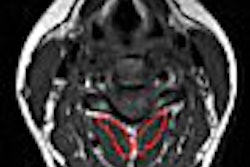
Using T1-weighted diffusion tensor MRI (DTI), Dr. Leonardo Bonilha and colleagues compared the brain scans of seven patients with dystonia with those of 10 patients without the disorder. They evaluated differences in mean diffusivity and fractional anisotropy, and found white-matter abnormalities in the patients with the dystonia.
In a poster presentation at the 2006 American Neurological Association (ANA) meeting in Chicago, Bonilha explained that the findings "suggest that idiopathic dystonia is not only a functional disorder, but it is also associated with structural brain changes. Impaired connectivity and disrupted flow of information may contribute to the impairment of motor planning and regulation in dystonia."
The researchers noted that idiopathic dystonia was associated with larger fractional anisotropy values in close proximity to the insula, putamen, claustrum, precentral gyrus, and cingulate and superior frontal gyri, according to the results.
They also saw smaller fractional anisotropy values in the pallidum, parahippocampal areas, thalamus, precentral and postcentral gyri, medial and middle frontal gyri, cingulate gyrus, precuneus, cuneus, and superior temporal sulcus, said Bonhila, who is an assistant professor of neurology at the university.
Finally, "idiopathic dystonia was also associated with increase in mean diffusivity in adjacent white matter to the pallidum and putamen bilaterally, left caudate, and in subcortical hemispheric regions including the postcentral gyrus, " he added.
To find ways to treat patients with idiopathic dystonia, it is imperative to first determine the cause of the condition and possibly where in the body the triggering genes reside so that pharmaceuticals can be directed at those regions or genes, Bonilha said.
"Abnormal fractional anisotropy and mean diffusivity in patients with idiopathic dystonia indicate that abnormal axonal coherence and integrity contribute to the pathophysiology of dystonia," he explained.
For more on the role of MRI in white-matter abnormalities, check out the following presentations at the 2006 RSNA meeting in Chicago:
- DTI in white-matter deficits for delayed cognitive impairments in patients who have suffered a head injury (scientific paper SSJ15-01)
- DTI in malformations of cortical development, such as transmantle focal cortical dysplasia and polymicrogyria (scientific paper SSK17-09)
- Optimizing fetal and neonatal MR for imaging normal and abnormal medullary vessels (education exhibit LL-PD5025)
- MR perfusion and DTI of microvascular white-matter disease (scientific paper SSQ13-08)
By Edward Susman
AuntMinnie.com contributing writer
November 24, 2006
Related Reading
Gray-matter abnormalities seen in autism spectrum disorder, August 22, 2006
MRI predicts neurodevelopmental outcomes in prematures, August 17, 2006
PET makes progress in documenting movement disorders, April 4, 2003
Copyright © 2006 AuntMinnie.com