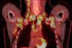
BOSTON - Researchers have found that FDG-PET and arterial spin labeling (ASL) MR perfusion match the gold standard of pathology or long-term clinical follow-up in differentiating tumor necrosis from recurrence in patients with brain tumors, according to a presentation at this week's American Roentgen Ray Society (ARRS) meeting.
Although both techniques demonstrated concordant results, ASL MRI may have an edge due to its quicker exam time compared to PET. Study results were presented Monday at the ARRS conference by co-author Dr. Fargol Booya of Beth Israel Deaconess Medical Center in Boston.
"One of the ways we have to distinguish radiation necrosis from recurrence is clinical follow-up, which, of course, is always too late, but it gives us the diagnosis," Booya said. "FDG-PET measures glucose uptake and is a marker of metabolic activity, while ASL measures blood flow, which is anticipated to be greater in areas of tumor recurrence."
Researchers analyzed 15 patients with 23 histologically proven brain tumors, who were treated with radiation or chemoradiation and had enhancing lesions at the site of primary tumor treatment in the follow-up imaging. Five of those patients had glioblastoma multiforme, and the other 10 patients had metastases.
Arterial spin labeling MRI and PET were performed within an average of 16 days of each other, with a maximum range of 50 days between procedures.
MRI versus PET
For each patient, the PET and ASL MRI studies were independently interpreted by a radiology resident and an attending neuroradiologist to determine if the patient had a tumor and to compare the lesions via both modalities. The readers then classified the lesion as either tumor recurrence or necrosis.
Standardized uptake values (SUVs) and perfusion levels of the lesion and the normal white matter were measured in FDG-PET and ASL studies, respectively. The ratio of FDG uptake of the lesion to normal white matter in PET and the ratio of cerebral blood perfusion of the lesion to normal white matter also were compared.
"Tumor recurrence, as we expected, initially showed higher FDG uptake of 2.9 [+ 0.8] and ASL perfusion ratio of 5.9 [+ 2.1]," Booya said. In addition, all tumor recurrences had SUVs greater than 5.7 and perfusion greater than 50 cc/min/g. In radiation necrosis, the uptake or perfusion was equal to or less than normal white matter, and reliable regions of interest could not be placed.
Close correlation
The researchers also found that no radiation necrosis had uptake greater than 4.9 SUV or a perfusion level greater than 29 cc/min/g. Booya noted a close correlation between FDG-PET findings and ASL in the tumor region of interest.
Using pathology or long-term clinical follow-up to confirm the PET and ASL findings, 11 lesions had 12 to 18 months of clinical follow-up, with 12 lesions having biopsy or resection.
In the final analysis, PET was concordant with pathology and/or long-term clinical follow-up in 18 of the 23 lesions, while ASL was concordant with pathology and/or long-term clinical follow-up in 22 of 23 lesions.
The results, Booya said, show that "ASL and FDG-PET are highly concordant with one another and the gold standard with pathological specimen and follow-up."
In cases where expediency is important, the researchers recommend the use of ASL, because its acquisition time of approximately five minutes is faster than a PET scan and "collecting perfusion data with ASL is a more efficient approach to answering a diagnostic question."
By Wayne Forrest
AuntMinnie.com staff writer
April 28, 2009
Related Reading
MRI, PET/CT help direct correct cervical cancer treatment, March 27, 2009
NOPR: PET changes care of more than a third of cancer patients, October 6, 2008
Diffusion-mapping MRI predicts radiation therapy response, April 22, 2008
MR perfusion imaging predicts malignant transformation of gliomas, March 27, 2008
PET/CT shows its worth in cervical carcinoma, January 18, 2008
Copyright © 2009 AuntMinnie.com