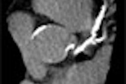
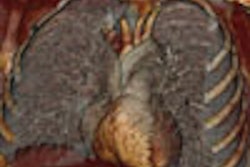
CHICAGO - FDG-PET/CT showed higher sensitivity than contrast-enhanced CT in the nodal staging of pulmonary lesions, according to information from an Italian research team. However, contrast-enhanced CT (CECT) revealed a higher specificity than FDG-PET/CT in characterizing pulmonary lesions, the group reported.
"The aim of our study was to compare fluorine-18 fluorodeoxyglucose PET/CT and contrast material-enhanced CT in the characterization and nodal staging of pulmonary lesions," said Dr. Emilio Quaia.
Quaia, from the department of radiology at the University of Trieste in Trieste, Italy, presented the results of his group's PET/CT and CECT research at the RSNA conference on Thursday.
The scientists retrospectively reviewed 76 consecutive patients, 56 males and 20 females with a mean age of 63.4 years. The patients presented with a total of 84 solid pulmonary lesions that ranged in size from 1 cm to 8 cm, according to Quaia. The lesions were identified primarily by plain film x-ray (72 total) or CT (12), he said.
The entire patient cohort had a CECT conducted on a Volume Zoom CT scanner (Siemens Medical Solutions, Erlangen, Germany), and also underwent a PET/CT study on a Discovery LS PET/CT system (GE Healthcare, Chalfont St. Giles, U.K.). The patients were injected with 370 MBq of F-18 FDG and had a nonenhanced spiral CT one hour after FDG intake, followed by a PET scan that covered the neck, thorax, abdomen, and pelvis, Quaia said.
Two independent panels of two readers each reviewed separated and fused FDG-PET/CT images and CECT images. The pulmonary lesions were characterized, by consensus, as malignant or benign according to a standardized uptake value (SUV) greater than 2.5 in the FDG-PET/CT images and reference standard criteria in the CECT images.
The gold standard of reference for final lesion characterization, according to Quaia, was histology from surgical or biopsied specimens.
"For nodal staging, histopathological diagnosis served as reference in 75 patients who underwent resective surgery with extensive N1 and N2 lymph nodes sampling or dissection," he said. "Surgical sampling included an average of six lymph node stations of the mediastinum."
The histopathological samples revealed a total of 72 malignant and 12 benign tumors, Quaia said. He reported that the sensitivity of FDG-PET/CT was 91.6% with a specificity of 16.6% in lesion characterization, while CECT demonstrated a sensitivity of 84.7% and a specificity of 58.3%. However, in nodal staging FDG-PET/CT showed a sensitivity of 76% and a specificity of 80%; CECT had a sensitivity of 48% and a specificity of 92%.
Quaia noted that FDG uptake in benign pulmonary tumors lowered the specificity outcome for FDG-PET/CT. He suggested that the research team may conduct further studies employing a dual-time-point FDG-PET protocol, which takes two series of PET images one hour apart, which he believes will improve the specificity of the modality.
By Jonathan S. Batchelor
AuntMinnie.com staff writer
December 2, 2005
Related Reading
PET/CT imaging useful in detecting and staging choroidal melanomas, November 8, 2005
Oncologic PET/CT: A primer for radiologists, October 14, 2005
FDG-PET/CT tops other technologies for lymphoma staging, March 23, 2005
PET/CT demonstrates staging strength over PET, CT, and PET plus CT, March 7, 2005
PET/CT hardware hybrid tops PET, software fusion for NSCLC staging, December 7, 2004
Copyright © 2005 AuntMinnie.com