CHICAGO - A significant number of patients with malignant melanoma of the skin initially staged as negative for lymph node and distant metastases with whole-body MRI and PET/CT eventually proved to have undetected regional or distant metastases, according to researchers from the department of radiology at the University Hospital of Essen in Germany.
"We conducted a prospective study to assess the accuracy of whole-body MRI in comparison with whole-body 18F-FDG-PET/CT for staging newly diagnosed malignant melanoma of the skin," said Dr. Florian Vogt, who presented the results of the team's study at the RSNA conference on Monday.
A total of 31 consecutive patients, 16 male and 15 female with a mean age of 45, underwent whole-body MRI on a 1.5-tesla Sonata system (Siemens Medical Solutions, Erlangen, Germany) and 18F-FDG-PET/CT imaging on a Siemens biograph PET/CT system for staging of lymph node (N) and distant (M) metastases after resection of the primary tumor, Vogt said.
The patients were imaged with an unenhanced T1- and T2-weighted HASTE sequence of the liver and thorax, followed by seven successive contrast-enhanced T1-weighted 3D VIBE sequences on the MR system.
Two radiologists evaluated the MR images, and a radiologist and a nuclear physician read the PET/CT images. The evaluation was done according to the American Joint Committee on Cancer Staging classification. Histology and a clinical follow-up (mean time 295 days) served as the standards of reference. Due to prior resection of the primary tumor, histopathology of all primary tumor sites was available, but T stage was not assessed separately, Vogt noted.
"Differences between the imaging procedures were tested for statistical significance by McNemar's test, and sensitivity and specificity values for both modalities were determined for N and M staging," Vogt said.
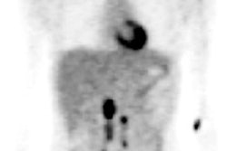
For N staging, PET/CT demonstrated a sensitivity of 50%, a specificity of 100%, a positive predictive value of 100%, a negative predictive value of 89.2%, and an accuracy of 90%. The MR imaging showed a sensitivity of 33.3%, a specificity of 100%, a positive predictive value of 100%, a negative predictive value of 86%, and an accuracy of 83%, Vogt reported.
For M staging, PET/CT imaging had a sensitivity of 37.5%, a specificity of 95.4%, a positive predictive value of 75%, a negative predictive value of 80%, and an accuracy of 77%. MRI demonstrated a sensitivity of 12.5%, a specificity of 95.5%, a positive predictive value of 76%, and an accuracy of 74%, according to the researchers.
Vogt admitted that the research group followed a strict protocol for determining false-negatives, on the basis of a clinical follow-up of approximately one year after imaging studies were preformed. However, the group believed that an N-stage or M-stage tumor probably was present at the time of the first imaging scan, he said.
"Based on the low sensitivities of staging newly diagnosed malignant melanoma of the skin, follow-up examinations -- either by whole-body MRI or PET/CT -- seem to be mandatory," Vogt concluded.
By Jonathan S.
Batchelor
AuntMinnie.com staff writer
November 30, 2004
Related Reading
PET better than standard methods at detecting recurrent melanoma, August 11, 2004
Radiation can control melanoma of the head, February 9, 2004
PET protocols from head to toe, May 5, 2003
Adequate follow-up contributes substantially to survival of melanoma patients, March 13, 2003
Copyright © 2004 AuntMinnie.com