In developing nations, cervical cancer remains a leading cause of cancer-related deaths, specifically because cytologic screening is not performed routinely. But approximately 30% of cervical cancers will recur, even if they are caught early with a Pap smear and subsequently treated with a multimodality protocol. A way to improve post-treatment surveillance for an earlier detection of relapse is necessary and 18F-FDG-PET may be the answer, according to a group from Taiwan.
Dr. Tzu-Chen Yen, Ph.D., and colleagues from Chang Gung University College of Medicine in Taoyuan set out to "define the prognostic features of recurrent cervical cancer patients for selecting appropriate candidates for (the) use of 18F-FDG-PET (Journal of Nuclear Medicine, October 2004, Vol. 45:10, pp. 1631-1639).
Yen's co-authors are from the departments of public health, ob/gyn, radiation oncology, and several other divisions of Chang Gung Memorial Hospital, also in Taoyuan. Additional contributors are from Taoyuan's Institute of Nuclear Energy Research and Mackay Memorial Hospital in Taipei.
The study involved 55 women who experienced treatment failure or unexplained elevated tumor marker serum levels, and proven relapse. Imaging was performed on an ECAT Exact HR+ PET system (Siemens Medical Solutions, Erlangen, Germany). The patients also underwent CT and MRI scans. The imaging protocols were detailed in a previous study authored by Yen's group (Journal of Clinical Oncology, October 2003, Vol. 21:19, pp. 3651-3658).
Three nuclear physicians interpreted the PET data with lesion status determined by pathology or clinical follow-up. The benefits of PET were calculated based on how the modality altered treatment.
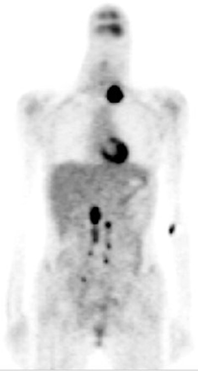
| A 25-year-old woman with poorly differentiated squamous cell carcinoma of the uterine cervix, FIGO stage 111b, underwent concurrent chemoradiation therapy. Three months later, a left neck mass was palpated. Abdominopelvic MRI and chest CT showed no definitely abnormal findings except an enlarged supraclavicular lymph node. |
 |
| In a mix of salvage radiotherapy and palliation treatment, PET was performed and suggested nodal metastases at the left supraclavicular, the bilateral upper and lower para-aortic, and the bilateral pelvic regions. After the left supraclavicular and para-aortic nodal metastases were confirmed histopathologically, patient received palliation treatment. Reprinted by permission of the Society of Nuclear Medicine from Yen TC, See LC, Chang TC, et al, "Defining the Priority of Using 18F-FDG-PET for Recurrent Cervical Cancer" (J Nucl Med 2004; 45: 1632-1639, p. 1634 [Figure 1]). |
According to the results, the interval between initial diagnosis of cervical cancer and the first document recurrence was four months to 23 years. In terms of the diagnostic efficacy of PET, the modality's sensitivity (89.2%) was significantly higher than CT and MRI (39.2%) for the detection of metastatic lesions. However, the difference was not noteworthy for local lesion detection (90% versus 80%).
Regarding treatment modification, 36 women (65.5%) had their treatment course altered because of PET findings. The significant factors associated with poor survival were primary radiation, squamous cell carcinoma antigen, and presence of symptoms at recurrence.
Using a prognostic scoring system, the women were divided into categories of low-, intermediate-, and high-risk groups. In the low-risk group, 10 patients had their treatment shifted to palliation from curative salvage therapy. In the intermediate group, a dozen also were switched to palliative treatment. In the high-risk group, five had their treatment changed post-PET.
By the end of July 2003, 18 patients had died with disease, 22 were alive with disease, and 15 were alive and disease-free, the authors stated.
"The benefits of PET are not only a reduction of mortality rates, but also avoidance of unnecessary salvage attempts (futile suffering and unrewarded medical cost)," the authors concluded. "We recommend that the priority use of PET in recurrent cervical cancer might be targeting those with a better prognosis to achieve maximal benefits."
While FDG-PET is still an expensive exam, the advantages in cervical cancer patients may outweigh the costs, suggested Dr.Tarik Belhocine, Ph.D., in an accompanying editorial.
"In the era of health cost-savings, 18F-FDG PET may be particularly useful by selecting the most efficient therapies in patients with good prognosis, and more so by avoiding unnecessary expenses in those of women with poor prognosis," Belhocine wrote (JNM, October 2004, Vol. 45:10, pp.1602-1604).
For more on the use of FDG-PET in cervical cancer, keep an eye out for the following presentations at the 2004 Radiological Society of North America (RSNA) meeting in Chicago:
PET Imaging in Radiation Treatment: Cervix I -- Technical, Refresher Course RC422B.
PET Imaging in Radiation Treatment: Cervix II -- Clinical, Refresher Course RC422C.
Uterine Cervical Cancer: Detection of Metastatic Lymph Nodes with Integrated FDG PET/CT, Scientific Session SSG25-06.
By Shalmali Pal
AuntMinnie.com staff writer
November 16, 2004
Related Reading
Demographics influence cervical cancer rates, July 28, 2004
Cervical cancer still cutting many lives short, April 22, 2004
Glucose metabolism regulators match FDG-PET uptake in cervical cancer, February 17, 2004
Copyright © 2004 AuntMinnie.com