SAN FRANCISCO - Assessing how much viable tumor remains after neoadjuvant therapy may be the best way to gauge treatment response. When it comes to soft tissue sarcoma, figuring out which patients will benefit from neoadjuvant therapy is an ongoing problem. Preoperative imaging with FDG-PET may offer a solution.
"People diagnosed with high-grade, localized soft tissue sarcoma (STS) have a high risk of developing metastasis after clinical resection and radiation therapy," said Dr. Scott Schuetze from the University of Washington in Seattle, in a presentation Tuesday at the American Society of Clinical Oncology meeting. "One of the problems with adjuvant chemotherapy is that only a minority of tumors are sensitive to currently available chemotherapy drugs."
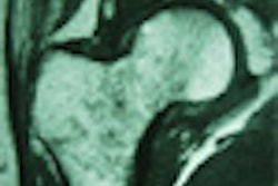
However, neither MRI nor CT has been particularly adept at revealing changes in the size and appearance of soft tissue sarcoma after treatment, mostly because both modalities are hampered by the presence of edema, inflammation, or extracellular material, Schuetze said.
Schuetze’s group tested the 18fluorodeoxyglucose (FDG) PET response to neoadjuvant chemotherapy. They also set out to see if that response could predict survival.
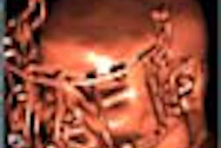
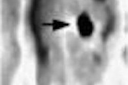
For the study, 56 patients with high-grade STS underwent FDG-PET imaging prior to, and after, 2-3 cycles of adriamycin-based chemotherapy. Imaging with the GE Advance PET scanner (GE Medical Systems, Waukesha, WI) commenced 45 minutes after injection of 7-10 mCi of FDG.
The acquired tumor image was corrected for single attenuation by normal tissues. A maximum standardized uptake value (SUV) for the tumor was calculated by drawing the region of interest around the tumor. Scans were obtained within one week of initiation of chemotherapy and surgery, Schuetze said.
"We decided to study the change in the maximum SUV for the tumor because many of the tumors showed a heterogeneous pattern of accumulation at the time of diagnosis, and a heterogeneous response to treatment. We reasoned that the most metabolically active portion of the tumor would dictate the biological aggressiveness of the tumor," he said.
The median pretreatment SUV was 5.6. After neoadjuvant chemotherapy and re-imaging with FDG-PET, the STS was resected. With a median follow-up of 22 months, 18 out of 56 patients developed local recurrence and 9 developed metastatic recurrence, Schuetze reported.
The median time to recurrence was 38 months in patients whose SUV was reduced by more than half, compared to 18 months for those with less than a 50% reduction in SUV. The median survival rate for the latter group of patients was 41 months. The median survival rate for the group with the higher SUV reduction has not been reached yet, Schuetze said.
"We find these preliminary results encouraging for the potential application of PET imaging as a method to predict the biological behavior of soft tissue sarcoma," he concluded. Future research will focus on finding the optimal time to perform FDG-PET imaging after treatment, and whether PET can help determine which patients will benefit from additional chemotherapy.
By Shalmali PalAuntMinnie.com staff writer
May 17, 2001
Related Reading
FDG-PET improves prognosis of esophageal cancer patients, May 15, 2001
FDG-PET proves its mettle in imaging cartilage lesions, May 2, 2001
Click here to post your comments about this story. Please include the headline of the article in your message.
Copyright © 2001 AuntMinnie.com