Concerns over security following the September 11 attacks caused a sharp drop in attendance at the 2001 RSNA meeting. But despite the thinner crowds, many radiology professionals who made the trip to Chicago said the quality of the meeting didn’t suffer. Clinical presentations went on as planned. Some imaging vendors even said the lower attendance made it easier to spend time with qualified equipment buyers.
On the clinical side, the most cutting-edge clinical presentations concentrated on areas such as computer-aided detection, multislice CT, digital radiography and mammography, virtual colonoscopy, and RIS/PACS integration.
Below is a short synopsis of meeting highlights, divided into categories based on the hottest trends at the show. The complete 2001 RNSA RadCast is available at http://radcast.auntminnie.com.
Computer-aided detection
Presentations on CAD showed that the new technology is proving itself to be a useful backup reader for mammographers.
Adding CAD to a double-reading mammography protocol can reduce false negatives by as much as 87%, according to a study by investigators at the Elizabeth Wende Breast Cancer Center in Rochester, NY. The researchers used CAD to examine mammograms with biopsy-proven cancers that were previously marked negative by mammographers.
Using CAD increased reading times to essentially that of a triple read, the investigators said. But another study, at George Washington University in Washington, DC, found that CAD can serve as an alternative to double reading. These researchers found that CAD increased a mammographer’s sensitivity by 21%, compared with the 5% to 15% increase generally associated with double reading.
CAD can be particularly useful for residents with limited experience in breast cancer detection, according to researchers at Necker Hospital in Paris. For a senior radiologist in the study, the CAD system led to a measurable increase in accuracy, from 76.9% to 84.6%, and found one missed cancer. But for the resident, CAD boosted accuracy from 61.9% to 84.6%, and the workstation found three cancers the junior radiologist missed.
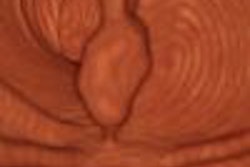
CAD is also moving into realms beyond breast imaging, with new applications being developed in lung nodule screening. CAD is also seeing rapid development in virtual colonoscopy. University of Chicago researchers presented their research on a CAD algorithm that analyzes shapes and textures in the colon to detect polyps. The researchers have developed additional techniques to reduce false positives. Other groups focused on specialized areas such as the detection of flat polyps.
Multislice CT
Multislice CT is another relatively new technology that is proving itself in the clinical setting. But the superb image quality and new applications enabled by MSCT have a downside: increased radiation dose.
Several RSNA studies addressed the radiation issue. Researchers at Duke University in Durham, NC, confirmed that MSCT delivers higher radiation dose if the same protocols used for single-slice scanning are employed.
The researchers used thermoluminescent radiation detectors placed in an anthropomorphic phantom that simulated areas of soft tissue, bone, and air space. They found that converting routine protocols from single-slice to multislice CT yielded a two- to-threefold increase in organ dose. The researchers urged multislice users to reduce mA wherever possible, and adjust protocols and parameters to maintain organ dose to levels equivalent to single-slice CT.
Another phantom study, by German researchers, found that multislice CT can deliver radiation doses in cardiac studies up to seven times higher than electron-beam CT, the current gold standard for CT cardiac imaging. They too recommended the development of optimized MSCT protocols.
A study from Massachusetts General Hospital in Boston demonstrated that it is possible to change protocols and cut mAs while maintaining diagnostic effectiveness. The group found that in noncritical applications such as routine follow-up exams, using half the radiation dose produced images that were just as useful as full-dose studies. While image quality was reduced, most structures remained well defined, and the technique was deemed acceptable in many circumstances for which higher-dose protocols are routinely used.
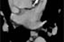
In the realm of new applications, multislice CT is having an impact on a number of cutting-edge procedures. One of them is coronary calcium screening -- the use of CT to detect calcium deposits in the heart that could be a precursor to a cardiac event.
MSCT can produce coronary calcium scores that are reliable and reproducible, according to a group from Johns Hopkins University in Baltimore. A group of readers from the institution were in close agreement in rating calcium scores, the researchers found. While variance of agreement increased as calcium scores rose, the presenter said that in any case, variances at the upper end wouldn't have a great effect on the course of therapy chosen.
Researchers at Children’s Memorial Hospital in Chicago found that dose reductions as low as 40 mAs for chest studies are possible without lowering diagnostic confidence. The technique reduced radiation exposure by an average of 66%, the researchers found.
A New York University group was able to reduce both image acquisition times and radiation dose in virtual colonoscopy by using MSCT rather than a single-slice scanner.
Another virtual colonoscopy study, by British researchers, indicated that virtual colonoscopy can be a tool with very high sensitivity and specificity for diagnosing colon cancer in symptomatic patients.
The investigators examined 1,400 patients with CTC over five years. They found 255 cancers with CTC, missing just 4 lesions that were detected with other modalities. Two of the missed cancers were flat lesions, while one was detected later with CTC when the bowel had been better prepared.
On the instrumentation side, imaging vendors pushed the envelope of CT technology, with several vendors showing works-in-progress 16-slice scanners, a major advance over the current four-slice machines. Look for most of these systems to be available later in 2002.
Digital radiography
Although the cost of the technology has limited DR’s widespread adoption, clinical studies are demonstrating an equivalence to film-screen radiography, and significant value in improving radiology department workflow.
A study presented in Chicago by German researchers focused on extremity imaging of the hand. A panel of judges rated DR as equivalent to or better than film-screen or CR, with DR scoring particularly well for border structures, soft tissue, and overall image quality. DR’s high dynamic range and linear characteristic curve enabled it to overcome low spatial resolution characteristics relative to film.
DR also got high marks in a study of chest images conducted by researchers at M.D. Anderson Cancer Center in Houston. Investigators reviewed images from 50 patients, taken with film-screen and DR techniques, and found that the readers preferred DR, especially for posteroanterior (PA) radiographs. DR also delivered lower patient radiation dose when equivalent speed settings were used.
Meanwhile, Japanese researchers found they could use DR to cut radiation dose without sacrificing image quality. Their study suggested that DR users could reduce dose to as low as a quarter of the yield in CR, depending on the thickness of the body part. And radiologists preferred DR images at half-dose to CR images at full-dose.
Digital mammography
Like digital radiography, full-field digital mammography is experiencing a slow adoption rate. But the sites that are using it appear to be pleased with the technology, according to several presentations.
A Norwegian study of 3,683 women compared FFDM with film-screen mammography. A total of 31 cancers were proven in the group, with the FFDM system finding 23 and film-screen 27. FFDM found three cancers that were undetected on film-screen, while film-screen found seven cancers that FFDM didn't see, although this result wasn't statistically significant (p=0.34).
Two of the four readers in the study had much higher false-negative rates with FFDM than with film-screen. The investigators said the higher rates were likely due to factors other than image quality, and that more training on FFDM was needed.
Other new technologies discussed at the meeting included breast MRI, breast imaging with through-wave transmission ultrasound, and radiolucent cushions to reduce pain caused by breast compression during exams.
RIS/PACS integration
Much of the work on IS/PACS integration is being carried out under the aegis of the Integrating the Healthcare Enterprise (IHE) initiative, a joint project between the RSNA and the Healthcare Information and Management Systems Society (HIMSS). The 2001 RSNA show marked IHE’s third year, and the IHE organizers chose to consolidate the gains of the past two years rather than roll out new integration profiles (the precise descriptions of how data standards like DICOM and HL7 are implemented).
Future work on IHE will create a general-purpose worklist capability, including tasks like review and reporting, as the next step beyond the current modality worklist, which is restricted to image acquisition. On the security side, the IHE committee is looking into whether to implement audit trails, and is also examining the development of a charge-posting feature.
Other PACS developments include Web-based software for distributing images to referring physicians in other specialties such as cardiology and orthopedics. Speech recognition and structured reporting applications also received attention, with several new companies debuting products to address these areas.
By Brian Casey
AuntMinnie.com staff writer
February 8, 2002
Copyright © 2002 AuntMinnie.com


