(Radiology Review) Radiologists from University of Michigan Hospitals in Ann Arbor have shown that using multidetector CT and fixed timing delays, maximal enhancement of vessels and hepatic parenchyma was achieved on either late arterial phase or portal venous phase imaging. They determined that in most cases, tumor detection was not improved during early arterial phase imaging, according to their paper in the American Journal of Roentgenology.
In order to determine which contrast-enhanced phase (early arterial, late arterial, or portal venous) displayed the best enhancement of the vessels and liver parenchyma, they examined 52 patients with suspected or known hepatic tumors. Also, they aimed to determine which contrast-enhanced phase enabled easiest detection of liver tumors.
"All patients were administered 150 mL of IV contrast material at an injection rate of 4 mL/sec. Images were acquired at 20 sec for the early arterial phase, 35 sec for the late arterial phase, and 60 sec for the portal venous phase," they reported.
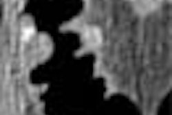
Seventy-three percent of hypovascular tumors showed improved or equivalent portal venous phase images compared with late arterial phase images, and for these tumors, portal venous phase images was better than early arterial phase images. Also, for the majority of hypervascular tumors, late arterial phase images were considered superior to early arterial phase images.
They reported, "because maximal celiac arterial and portal venous opacification is obtained in the late arterial and portal venous phases, respectively, and because the use of early arterial phase images does not contribute to tumor detection in most patients, we no longer routinely perform early arterial phase imaging in patients with known or suspected hepatic neoplasms."
"In summary, although we found that late arterial and portal venous phase images had better multidetector CT results than early arterial phase images for hypervascular liver tumors, this finding was not statistically significant," they wrote. "For hypovascular liver tumors, late arterial and portal venous phase images had statistically significant superior maximal tumor-to-parenchyma differences compared with early arterial phase images," they concluded.
Multidetector CT of the liver and hepatic neoplasms: effect of multiphasic imaging on tumor conspicuity and vascular enhancement Francis, I. R. et. al.Department of radiology, University of Michigan Hospitals, Ann Arbor, MI
AJR 2003 May; 180:1217-1224
By Radiology Review
August 18, 2003
Copyright © 2003 AuntMinnie.com