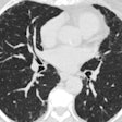
By any measure, CT pulmonary angiography (CTPA) is one of the bright lights of the decade in medicine. The development and continuous evolution of multidetector-row CT scanners have increased accuracy of the exam to 90% and higher for detecting pulmonary embolism (PE), triaging patients in the emergency room, and ultimately sparing lives.
In the small percentage of CTPA findings that are indeterminate, however, motion artifacts and contrast delivery are usually to blame, researchers from Hôpital Bichat in Paris concluded in the October issue of Radiology (October 2005, Vol. 237:1, pp. 329-337).
"With the high sensitivity (as much as 94%-96%) and specificity (as high as 94%-100%) of recently developed CT pulmonary angiography techniques, treatment protocols resulting from a diagnostic study are well-defined: A positive study results in anticoagulation treatment or placement of vena cava filter, while a negative study necessitates an alternative diagnosis (which is often supplied as an incidental finding from chest CT)," Dr. Stephen E. Jones, Ph.D., and Dr. Conrad Wittram wrote, noting that clinical outcomes from indeterminate studies are not well-described.
The Paris study took a retrospective look at 3,612 CTPA reports over a two-year period -- following a keyword search for "indeterminate," "nondiagnostic," or "inadequate" on the hospital database, yielding a match in the reports of 237 patients (mean age 57; 117 men, 120 women) whose exams had failed. In addition, 25 randomly selected diagnostic studies (mean age 64; 117 men, 120 women) were used as a control group.
"The percentage of 'indeterminate' studies as reported in articles on 10 studies ranges from 0.5% to 10.8%, with a mean of 6.4%," the researchers noted. "To our knowledge, the causes and proportions for indeterminate studies have not previously been systematically analyzed."
All images were acquired in a craniocaudal direction on either a four- or 16-detector-row scanner (GE Healthcare, Chalfont St. Giles, U.K.) in a single breath-hold. The institution's protocol called for spiral scanning from the lung apices to the adrenal glands at 380 mAs, following the injection of 135 mL of iopamidol (Isovue 300, Bracco, Milan, Italy) at 4 mL/sec.
Scanning was started after the contrast was maximally distributed in the pulmonary arteries, generally 20 to 30 seconds after injection depending on the body habitus of the patient, the authors wrote. Automated dose reduction was not used. Images were reviewed in on an IMPAX workstation (version 4.1, Agfa HealthCare, Mortsel, Belgium) using PE settings.
Both diagnostic and nondiagnostic studies were reviewed for the use of follow-up imaging, including repeat CTPA, conventional pulmonary angiography, ventilation-perfusion scintigraphy, or lower-extremity ultrasonography, as well as the use of anticoagulants and vena cava filter placement. The reports were also reviewed for patient outcomes and comments regarding the indeterminate nature of the CTPA exams. "Studies (in patients and control subjects) were reviewed for PE, contrast attenuation in the main pulmonary artery (MPA), motion artifacts, image noise, and flow artifacts," the group wrote.
"The cause cited for indeterminism was most often motion (74%) followed by poor contrast enhancement (40%)," the team found. Beam-hardening artifact and parenchymal disease were also cited, but infrequently. In all, 77 (32%) of the studies that met minimally acceptable motion and bolus enhancement standards were still read as indeterminate.
Contrast attenuation, as assessed by the measure of mean Hounsfield units and standard deviation (SD) in the main pulmonary artery, was 245 HU ± 80 SD in the patients, and 339 HU ± 88 in the control subjects. Just 46% of the indeterminate studies met the institution's criteria for adequate attenuation in the MPA.
"The assessment of poor bolus enhancement is largely unaffected by the presence of motion, whereas poor bolus enhancement was most likely inadequate timing of bolus administration," the authors reported.
Five PEs (2.1%) were found that had been missed in the original studies, two caused by imaging artifact. A transient lack of contrast was noted on 10 studies.
"Significantly delayed bolus timing, as indicated by the lack of contrast enhancement in the superior vena cava, was identified on five studies," the group wrote. "Conversely, on three studies there was poor vena cava enhancement caused by superior vena cava stenosis."
A persistent problem with CTPA studies is that patients tend to be anxious, dyspneic, and therefore often unable to hold their breath for the length of the scan, the team wrote. Also tricky is the bolus timing -- delivery too early or too late can render an indeterminate study. Mistiming can result from technician error, anatomic variations, or, most commonly, differences in cardiac output, the group explained.
"Our results show that 6.6% of CT pulmonary angiograms are indeterminate, mostly because of respiratory motion, followed by poor bolus enhancement," Jones and Wittram concluded. "The radiologist has a lower threshold for the presence of motion effects than for poor bolus enhancement."
By Eric Barnes
AuntMinnie.com staff writer
October 19, 2005
Related Reading
Triage with chest x-ray expedites PE evaluation, September 16, 2005
Widen that search: CTPA reveals more non-PE than PE diagnoses, May 18, 2005
Meta-analysis shows CT is sufficient to exclude PE, April 27, 2005
Multidetector CT does not improve outcomes of suspected PE, April 12, 2005
CT venography plus CTPA finds more pulmonary embolism, February 1, 2005
Copyright © 2005 AuntMinnie.com


